|
Abstract:
The growing need of various orthopaedic procedure as a part of
treatment of above pathologies demands accurate knowledge of
measurements of the vertebra. The aim of our study is to
determine the vertical and transversel diameters of pedicles and
to have a morphological database of the vertebral pedicles,To
compare and correlate this study with the available data,To
discuss the various parameters with respect to applied anatomy.
From our study we concluded that there is always an increase in
width of lumbar pedicles proceeding from L1 to L5 levels and
width being maximum at L5 level to enable in weight
transmission.
J.Orthopaedics 2009;6(4)e9
Keywords:
Cadaveric study; vertebral pedicle; Transvers diameter; Vertical
diameter.
Introduction:
Measurement of bone or any part of human body provides accurate
knowledge about morphology of the structure which helps the
clinicians, in diagnosing and treating various diseases. Due to
present lifestyle and with its speed, it has resulted in
increase in the incidence of assaults on the vertebral column in
the form of different spinal pathologies such as prolapsed inter
vertebral discs, spondylolisthesis, spondylosis, fractures. The
growing need of various orthopaedic procedure as a part of
treatment of above pathologies demands accurate knowledge of
measurements of the vertebra. The present study was undertaken
with the view to study lumbar region pedicles. Posterior spinal
instrumentation is a time honored method of spinal fixation.
Transpedicular fixation of the spine with pedicular screws is
becoming increasingly popular as it is more stable and versatile
because it provides three dimensional fixations. Several systems
of internal fixation that uses the pedicle as a source of
purchase for bone screws from posterior approach into the
vertebral body are currently available. These systems depend on
the ability of screws traversing the pedicles, the strongest
part of vertebrae even in severe osteoporosis, to hold in until
solid fusion occurs.
In several studies, researchers demonstrated fusion rates of 90%
or greater with pedicle screw fixation. However along with this
benefit a number of complications associated with pedicle screw
fixation were reported. The most devasting complication related
to pedicle screw is neurological injury secondary to misplaced
screw abutting or transecting a nerve. So with the use of
pedicle screw system it becomes imperative that a causal
relationship between the screw and neurological complication be
ruled out.
Morphometric study of pedicles of spine of dorsal and lumbar
region is thus relevant and critical for proper placement of the
traspedicular screw to avoid inadvertent penetration of
pedicular wall. Accurate anatomical description of the shape and
orientation of dorsal and lumbar pedicles are necessary for use
of implantable devices and spinal instrumentation techniques.
Internal fixation methods are increasingly used for lateral
fusion after a wide midline decompression, especially if
articular processes have been scarified. Several studies have
investigated the morphometry of thoracic pedicles with the use
of various techniques such as direct measurements and on plain
radiographs or computerized tomographic scans.
It is important to distinguish differences in the morphometry of
dorsal and lumbar pedicles. Studies were conducted where data
have been obtained from radiographs of spines and then compared
with direct measurements. These data were then used to verify
the accuracy of the technique that radiologist and surgeons must
use for pre-operative evaluatio.
The aims of the present study are:-
1.
To determine the vertical and transversel diameters of
pedicles and to have a morphological database of the vertebral
pedicles.
2.
To compare and correlate this study with the available
data.
3.
To discuss the various parameters with respect to applied
anatomy.
Importance and application:-
Morphologic data base will help in the standardization of the
dimensions of the pedicles of vertebrae in Indian population.
There has been growing interest in internal spinal fixation.
Pedicle is important site for same .This anatomic database will
definitely reduce the disparity between the pedicle screw size
and the pedicle. Pedicle diameters are large enough in most
cases to allow substantially larger screw diameter than
currently used. This would improve the strength of transpedicle
screw fixation. It will be helpful in designing of the spinal
instruments.
Data can be useful while carrying out surgical procedures with
the help of model vertebral column in the operation theatre.
Materials
and Methods:
This is an observational study.
The study was carried out in the Department of Orthopaedics and
Department of Anatomy, Grant Medical College and Sir J.J. group
of Hospitals, Mumbai.
For this study, 25 cadavers, ranging from 2 months to 24 months
post embalmed were selected from the Department of Anatomy,
Grant Medical College and Sir J.J. group of Hospitals, Mumbai.
Cadavers showing obvious deformity in the spine or a fractured
spine were excluded from this study.
Cadavers were dissected using the midline posterior approach so
as to expose the spine upto the sacrum. The facet joints and the
vertical bony crest just below it were cleared of the soft
tissue.
Direct measurements were taken on the vertebra with using a
sliding vernier caliper and divider.
Vertical Height of pedicle:
It was noted by a sliding vernier caliper and divider. The
points just opposite each other on the upper and lower margins
of pedicles, in the vertical plane on its lateral aspect, where
the diameter was maximum were considered. First record was taken
on right pedicle and then on left.
Pedicle Width:
The deepest point on the lateral and medial aspect of each
pedicle were chosen. The thickness was measured at these points,
at right angles to the long axis of pedicle. First reading was
taken for right pedicle and then for the left.
Results :
There were 25 cadavers selected from department of anatomy for
this study.
The cadavers showing obvious deformities in the spine or a
fractured spine were excluded from this study.
Transverse pedicle
diameter
The transverse diameter increased gradually from L1 to L5.
The maximum transverse diameter was found at L5 level.
The maximum mean transverse diameter was found at L5 (12.001
mm).
The range of maximum transverse diameter was from 9 to 20 mm at
L5 level.
The minimum mean width of pedicle was found at L1 and was 7.242
mm.
The range of transverse diameter at L1 was from 5 to 11 mm.
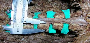
Fig 1:Transverse pedicle diameter.
Vertical pedicle
diameter
The vertical pedicle diameter decreased marginally from L1 to L3
but again increased at L4 and abruptly increased at L5.
The minimum vertical pedicle diameter was noted at L3.
The range of vertical pedicle diameter atL3 was from 12 to 16 mm
and mean at L3 level was 14.167 mm.
The maximum vertical pedicle diameter was recorded at L5.
Range of vertical pedicle diameter at L5 was from 14 to 25 mm
and mean at L5 was 19.22 mm.
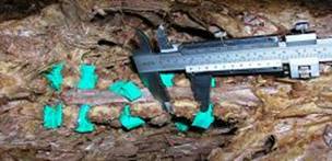
Fig 2: Vertical pedicle diameter.
Measurement of lumbar pedicle transverse diameters
|
LEVEL |
|
PDW |
|
L1 |
MEAN |
7.242 |
|
S.ERROR |
0.21 |
|
RANGE |
5 -11 |
|
STD. DEVIATION |
2.22 |
|
L2 |
MEAN |
7.864 |
|
S.ERROR |
0.25 |
|
RANGE |
5 – 12 |
|
STD. DEVIATION |
2.66 |
|
L3 |
MEAN |
9.106 |
|
S.ERROR |
0.26 |
|
RANGE |
6.5 – 14.5 |
|
STD. DEVIATION |
2.79 |
|
L4 |
MEAN |
10.45 |
|
S.ERROR |
0.22 |
|
RANGE |
7.5 – 13 |
|
STD. DEVIATION |
2.35 |
|
L5 |
MEAN |
12.001 |
|
S.ERROR |
0.41 |
|
RANGE |
9 – 20 |
|
STD. DEVIATION |
4.39 |
Measurement of lumbar pedicle vertical diameters
|
LEVEL |
|
PDH |
|
L1 |
MEAN |
14.227 |
|
S.ERROR |
0.17 |
|
RANGE |
11 – 16.5 |
|
STD. DEVIATION |
1.86 |
|
L2 |
MEAN |
14.227 |
|
S.ERROR |
0.17 |
|
RANGE |
12 - 18 |
|
STD. DEVIATION |
1.84 |
|
L3 |
MEAN |
14.167 |
|
S.ERROR |
0.15 |
|
RANGE |
12 – 16 |
|
STD. DEVIATION |
1.57 |
|
L4 |
MEAN |
14.909 |
|
S.ERROR |
0.23 |
|
RANGE |
12 – 19 |
|
STD. DEVIATION |
2.41 |
|
L5 |
MEAN |
19.227 |
|
S.ERROR |
0.38 |
|
RANGE |
14 – 25 |
|
STD. DEVIATION |
4.03 |
Discussion:
Studies have been already conducted in white and a few
non-whites population. However, only very few studies have been
conducted in Indian population. Moreover, many studies have not
reported all the morphometric dimensions related to pedicle
screw placement.
If the dimensions of pedicles change at each vertebral level,
information on this might help to prevent failure of fixation
and injury to surrounding vital structures.
From above discussion, it is apparent that dimensions of
pedicles of lumbar spine play a crucial role in success rate of
pedicle screw fixation. The knowledge of pedicle morphology is
essential for proper placement of the pedicle screws. Analysis
of pedicle dimensions can be obtained from direct measurements,
radiographs, computerized tomographic scans.
Berry-(1987) (1)
- Studied selected thoracic and lumbar vertebrae. Total 30
vertebral columns were studied. He restricted his work to T2,T7,
T12 and L1 to L5. He used the vernier caliper and the outside
dimension Caliper and the angular measurements were recorded
with a goniometer.
Zindrick(1987)
- Study was carried out on the lumbar vertebrae from 170
vertebral columns. Total 2905 pedicles were measured.
Measurements were made from the individual vertebral specimen
roentrogram.
Krag (1988)
– Mainly studied pedicles in the lower thoracic and lumbar
region. This was actually a retrospective review of the C.T.
Scans of the thoracic and the lumbar spines. Thus the X-ray
imaging technique was used for the study.
Scoles(1988)
– He employed the resources of the Hamman and Todd Osteological
Collection at the Cleveland Museum of Natural History. They
studied selected thoracic and lumbar vertebrae. The number of
vertebral column studied was fifty. He observed that the
posterior element morphology is highly variable and largely
unpredictable. Linear measurements were made with the vernier
caliper and the protractor. Angular measurements were taken with
the goniometer. All the measurements were taken directly from
the bony specimens. They calculated the mean, standard
deviation, maximum and the minimum values for each. The linear
regression analysis was used to search for co-relation between
the measurements and also between the individual anthropometric
data and the vertebral measurements.
Olsewski(1990)
- Lumbosacral spines from 49 emblamed cadaver were used for the
study. The pedicles of lumbar vertebrae were measured both
directly and radiographically. The study was divided into 3
parts : -
1.
The direct measurement of lumbar pedicles in cadavera.
2.
The comparison of measurements on radiographs of lumbar
pedicles in cadavera with direct measurements
3.
The measurements of lumbar pedicles on radiographs and
computerized tomographic scans of living young adults patients.
Panjabi (1991)
- Study was carried out on the fresh autopsy specimens. He
studied 144 thoracic vertebrae with the help of a three
dimensional morphometer. The vertebra was secured in a
morphometer stand and then subjected to the measurements, the
linear as well as angular, by the three dimensional mophometer.
Amonoo –Kuofi(1995)
– Has reported on his study of horizontal and vertical diameters
of pedicle of lumbar verterbrae. Study was done on plain
radiographs and measurements of lumbar pedicle were measured
using a vernier caliper.
Sajal Mitra(2002)
- Studied the morphometry of lumbar pedicle in Indian population
as related to pedicle screw fixation. 20 cadavers were dissected
and the measurements of lumbar pedicles were measured using a
vernier caliper.
Singel T C(2004)
– Study was conducted on 60 adult lumbar vertebrae. Direct
measurements of horizontal and vertical diameter of pedicles of
lumbar vertebrae were done on the preserved set of bones of
individual dead bodies. Measurements were done by vernier
caliper.
From above discussion, it is apparent that dimensions of
pedicles of lumbar spine play a crucial role in success rate of
pedicle screw fixation. The knowledge of pedicle morphology is
essential for proper placement of the pedicle screws. Analysis
of pedicle dimensions can be obtained from direct measurements,
radiographs, computerized tomographic scans.
Conclusion:
According to the above discussion, the present study concludes
that there is always an increase in width of lumbar pedicles
proceeding from L1 to L5 levels and width being maximum at L5
level to enable in weight transmission.
Referring to the above discussion, the present study concludes
that the height of lumbar pedicles decreases as we move from L1
to L3 and again increases at L4 and maximum at L5.
The lordotic lumbar curvature of the vertebral column affects
the pedicle which show a splaying effect. This is the result of
the posterior shift of the weight transmission line.The splaying
and tilting was obviously found in the pedicles of L4 and L5
vertebrae in present study. So one has to be aware of the
misinterpretation of values of maximum and minimum pedicle
diameters while reading computerized tomographic scans. So it is
suggested that the patient should be given a proper position to
get the exact minimum and maximum pedicle diameters at these
levels.
Screw fixation of pedicle is safe method provided the transverse
cortical diameter of the pedicle is accurately determined.
To avoid any untoward complication, a image intensifier should
be used when inserting transpediclualr screws, particularly at
L5.
Reference :
-
Berry JL, Moran JM, Berg WS et al (1987) : A morphometric
study of human lumbar and selected thoracic vertebrae. Spine;
12:362-366.
-
Zindrick MR, Wiltse LL, Doornik A, et. al., (1987) : Analysis
of the morphometric characteristics of the thoracic and lumbar
pedicles. Spine vol.12; 160 – 166.
-
Krag MH, Weaver DL, Beynon BD (1988) : Morphometry of the
thoracic and the lumbar spine related to transpedicular screw
placement for surgical spinal fixation. Spine; 13 : 27 – 32
-
Scoles PV, Linton AE, Latimer B, Levy ME, Digiovanni BF (1988)
: Vertebral body and posterior element morphology: The normal
spine in middle life. Spine Vol.13 : 1082 – 1086.
-
Olsewski JM, Simmons EH, Kallen FC et. al., (1990) :
Morphometry of the lumbar spine : Anatomical perspectives
related to transpedicular screw fixation. JBJS 1990 : 541 –
549.
-
Panjabi M, Takata K, Goel V et. al.,(1991) : Thoracic human
vertebrae, Quantitative three dimensional anatomy. Spine 16 :
888 – 901.
-
Sajal Mithra, Datir SP et. al., (2002) : Morphometric study of
lumbar pedicle in the Indian population as related to
pedicular screw fixation. Spine Vol.27 ; 453 – 459.
-
Singel TC, et. al., (2004) : A study of width and height of
lumbar pedicles in saurashtra region. J. Anat. Soc. India 53;
4.
|

