|
Tsuyoshi Nakai* and Masaaki Kakiuchi **
*Department of Orthopaedic Surgery, Itami City Hospital
** Department of Orthopaedic Surgery, Osaka Police Hospital
Address for Correspondence:
Tsuyoshi Nakai
Department of Orthopaedic Surgery, Itami City Hospital
1-100 Koyaike, Itami City, Hyogo, 664-8540, Japan.
Phone: 81-72-777-3773
Fax : 81-72-781-9888
E-mail:
tsuyoshi223@gmail.com
|
|
Abstract:
An
anterolateral approach is in use for minimally invasive total
hip arthroplasty. This approach uses an intermuscular plane
between the tensor fasciae lata and gluteus medius. To our
knowledge, although a technique has been reported in a lateral
decubitus position, no report has been made in a supine position
on a standard operating table. We describe a minimally invasive
total hip arthroplasty technique done through an anterolateral
approach on a supine position.
J.Orthopaedics 2009;6(3)e10
Keywords:
minimally invasive total hip arthroplasty; supine position;
anterolateral approach; instrument
Introduction:
Total
hip arthroplasty (THA) remains one of the most successful
surgeries done by orthopaedic surgeons through a variety of
surgical approaches. Recently, minimally invasive hip
arthroplasties have been in popularity, done with a variety of
surgical approaches (1,2,3,4). Surgical approaches and implant
positioning have been recognized as factors influencing hip
arthroplasty stability. There is some agreement that minimally
invasive approach refers to minimizing soft tissue damage
during THA. Kim and Heinz reported anterolateral mini-incision
hip replacement surgery in a lateral decubitus position which
the posterior capsule intact so that posterior dislocation could
be minimized (5). However, it is still difficult to check leg
length discrepancy because of a lateral decubitus position. The
purpose of this article is to describe in detail anterolateral
approach using a standard operating room table on a supine
position.
Technical note:
The anterolateral approach for total hip arthroplasty is
performed in a supine position on a standard operating room
table. The perineum is placed at the break in the foot of the
bed so that when the foot of the bed is lowered, hip extension
will result. Before draping, the nonoperative hip was placed in
abduction position on the table. This will later allow more
adduction of the operative leg. Pneumatic compression boots
were applied to nonoperative leg for intraoperative deep venous
thrombosis prophylaxis. The operative leg is completely draped
free into the field to allow full range of motion of the hip
during the surgery. Preoperative templating of radiographs gave
an initial plan for acetabular shell size, level of neck cut,
femoral stem size, and head-neck length. The anterolateral
approach is a modification of the Watson-Jones approach using
the proximal portion. An incision is made that begins 3 to 4 cm
posterior to the anterior superior iliac spine. The incision is
the extended slightly obliquely in a posterior direction for 8
to 10cm. Dissection is carried out down in the Watson-Jones
interval. This anterolateral approach to THA, which exploits
the interval the tensor fasciae lata and gluteus medius muscle
for both acetabular and femoral preparation, allows for primary
exposure of the hip joint capsule without any muscle damage.
The tensor fasciae lata, rectus femoris, and gluteus medius are
retracted, but not cut. The anterior hip capsule can be
excised. After releasing the hip capsule, the femoral neck
osteotomy, and removing the femoral head was made. Two tined
retractor can be placed outside the acetabular labrum
posteriorly, but inside the hip capsule and around the posterior
acetabulum. This two-tined retractor has made of different
length with 5mm and 10mm tines. Two versions of this retractor
for the left and the right hip exist (Fig). For left side hip
retractor, left side tine is 5mm, and right side tine is 10mm,
which facilitate to put the retractor to the posterior
acetabulum. A blunt tipped cobra retractor can then be placed
just anterior to the anterior portion of the acetabular fossa
around the anteroinferior acetabular wall. A sharp tipped
Hohmann retractor can be placed anterior acetabular wall, in a
position that puts it roughly perpendicular to the ilioinguinal
ligament. Thus, view and access to the acetabulum seem to be
equal to any other approach for hip arthroplasty. Cemented as
well as uncemented components have been placed through this
approach depending on surgeon preference. The legs are now
positioned for femoral preparation. The nonoperative leg is
placed in abduction. The operative leg is hyperextended,
slightly adducted, and externally rotated. We use an orthpaedic
table that the leg support can be broken and flexed independent
of the part of the table supporting the patients torso. It is
important throughout the femoral portion of the procedure to
keep the knee straight. Flexing the knee will cause increased
tension on the rectus femoris muscle and tends to drive the
proximal portion of the femur posteriorly, decreasing the
exposure. A two pronged retractor with relatively short tines
is then placed on the posteromedial calcar region. A second two
tined retractor, with relatively long tines, is then placed
around the outside the greater trochanter. It is important to
place this retractor between the greater trochanter and the hip
abductors, but outside the superior hip capsule. The proximal
femur is then pulled anteriorly and laterally with a small bone
hook inside the calcar. It is necessary to release superior and
posterosuperior capsule at least all the way back to the
posterosuperior corner of the femoral neck. To minimize the
need for elevation of the femur, we use a double offset broach
handle with lateral and anterior offset (Fig). After broaching
is complete, the trial femoral neck and head can be placed.
Stability and range of motion can be completely checked easily
because the operative leg is draped free. Leg length can also
be checked at the medial malleoli because both are readily
palpable in the supine position. Cemented as well as uncemented
components have been placed through this approach depending on
surgeon preference.
In
summary, minimally invasive anterolateral total hip arthroplasty
can be performing safely and precisely on a supine position on a
standard operating table using a two tined retractor and double
offset broach.
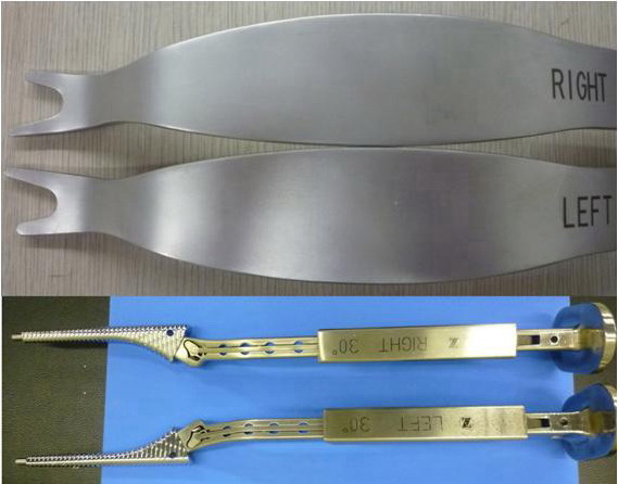
Fig:
Two versions of retractor exist for the left and the right hip,
for left side hip retractor, left side tine is 5mm, and right
side tine is 10mm (lower part).
Double offset broach handle with lateral and anterior offset
(upper part).
Reference :
-
Bal BS,
Haltom D, Aleto T, Barrett M. Early complications of primary
total hip replacement performed with a two-incision minimally
invasive technique. Journal of Bone Joint Surgery (Am )
2005; 87: 2432-2438.
-
DiGioia
AM
Ⅲ,
Plakseychuk AY, Levision TJ, Jaramaz B. Mini-incision
technique for total hip arthroplasty with navigation. Journal
of Arthroplasty 2003; 18: 123-128.
-
Wenz JF,
Gurkan I, Jibodh SR. Mini-incision total hip arthroplasty: A
comparative assessment of perioperative outcomes. Orthopedics
2002; 25: 1031-1043.
-
Wright
JM, Crockett HC, Delgado S, Lyman S, Madsen M, Sculco TP.
Mini-incision for total hip arthroplasty: A prospective,
controlled investigation with 5-year follow-up evaluation.
Journal of Arthroplasty 2004; 19: 538-545.
-
Kim CB,
Heinz R. Anterolateral mini-incision hip replacement surgery:
A modified Watson-Jones approach. Clinical Orthopaedics and
Related Res earch 2004; 429: 248-255.
|

