|
Abstract:
Tendon dislocations around the ankle joint are usually occurred
during the sports injuries. The traumatic posterior tibial
tendon dislocation is rarely reported, however spontaneous
dislocation is never reported in the English literature. Authors
reports a case of non-traumatic dislocation of posterior tibial
tendon and clinical result
Keywords:
posterior tibial tendon; dislocation; non-traumatic
J.Orthopaedics 2007;4(4)e15
index.htm
Introduction:
Dislocation of the posterior tibial tendon is a rarely reported
injury in the English literature. Most of the previous reports
had been neglected chronic dislocation of the posterior tibial
tendon. The cause is commonly related to trauma with an
inversion and dorsiflexion of the ankle and disruption of the
retinaculum.
In this report, we address a case of acute, non-traumatic
dislocation of the posterior tibial tendon in a 48-years-old
plumber.
Case Report :
A 48-years-old,
male plumber was referred for pain and swelling over the medial
side of the ankle. He denied any previous episode of ankle
injury or instability. He had no history of diabetes or
neruomuscular disorders.
On the day the
patient was referred to our facility, he woke up and walked
several steps. He felt sudden muscle clamping pain on his right
calf, and in attempts to relieve this, he stamped his foot on
the floor and twisted his ankle manually. He then experienced
swelling on the medial side of the ankle with pain.
Physical
examination demonstrated edema and tenderness around the medial
malleolus. The patient was able to walk but was unable to push
up into a tip toe position. The patient felt pain while
resisted invert his plantarflexed foot and was apprehensive when
the posterior tibial tendon was forced to push anteriorly.
A plain radiograph
of the ankle joint revealed no abnormality. Magnetic
resonance(MR) imaging of the ankle in a neutral position showed
anterior subluxation of posterior tibial tendon and surrounding
effusion in the transverse plane.
The posterior
tibial tendon was found to be properly intact with continuity
and showed no evidence of tendon degeneration(Fig.1).
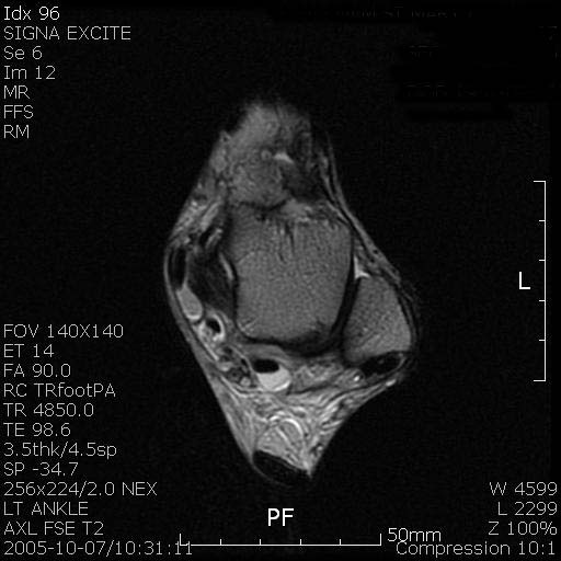
Fig. 1 .MR image shows tenosynovitis of posterior tibial
tendon and surrounding structures.
The patient underwent exploratory surgery. Under the general
anesthesia, the toruniquet was applied with the patient in the
supine position. The posterior tibial tendon was easily
dislocated manually over the medial malleolus by pushing it
anteromedially.
A 5cm long longitudinal skin incision was made along the course
of posterior tibial tendon at the posterior border of tibia.
There was evidence of acute tenosynovitis of the posterior
tibial tendon with increased synovial fluid.
The retinaculum was normal in thickness and minimally stretched
but had no pseudocapsule on the surface of the tibia.
The retinaculum was
incised longitudinally and focal ecchymosis of inner surface of
retinaculum was noticed(fig.2). The inflamed synovium was
excised. The posterior tibial tendon appeared to be normal in
shape. The periosteum was elevated and posterior tibial cortex
was exposed.
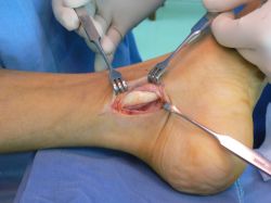
Fig.2. Posterior tibial tendon is normal in thickness
and inner surface of retinaculum had focal ecchymosis.
A 2cm long and 1cm wide trapdoor was made using a
micro-oscillating saw and the fragment was transpositioned 5mm
posteriorly to deepen the groove. The fragment was fixed with 2
screws and the retinaculum was sutured using absorbable suture
material.
The posterior tibial tendon was noted to glide smoothly with no
dislocation or 5mm subluxation for the full range of ankle
motion.
Postoperatively the ankle was placed in a fiber-glass cast for 4
weeks with slight plantarflexion and inversion. Partial weight
bearing ambulation was allowed by walking with the aid of
crutches on the day of surgery and during the patient return
home.
A gentle active range of motion was initiated and full weight
bearing walking ambulation was permitted at 6 weeks
postoperatively.
The patient returned to full physical activity at 12 weeks
postoperatively.
At the most recent follow-up of 18 months, the patient had no
symptom related to the ankle with normal range of motion and
returned to pre-injury level of activity without recurrence of
dislocation.
Dsicussion:
Compared to dislocation of the peroneal tendon, posterior tibial
tendon dislocation is a rare injury. The most common injury is a
trauma injury during the sports activities or dancing and the
proposed causative mechanism is a forced dorsiflexion with
inversion and a sudden tensioning of the posterior tibial
tendon(1).
In the case of our patient, he had no ankle injury history. The
patient had a sudden muscle cramp and swelling around the medial
malleolus in the morning.
In the operative field, the retinaculum appeared to be
over-stretched and focal ecchymosis was found on the inner layer
of the retinaculum and the posterior tibial tendon sheath. The
posterior tibial tendon was appeared to be nomal in thickness
and had a slight focal color change which may have resulted from
trauma during dislocation. The groove for the posterior tibial
tendon was normal in depth, however, the tendon was easily
dislocated anteriorly with finger pressure.
Reported operative treatments have been similar to dislocation
of the peroneal tendon which include reconstuction of the
retinaculum or deepening of the groove.
Reconstruction of the retinaculum consists of reattaching the
periosteal-retinacular sleeve to the anatomical margin of the
groove and most reports of this procedure were satisfactory for
the traumatic dislocation of the posterior tibial tendon(2,3)
In our patient, the dislocation was non-traumatic and the
retinaculum did not appear over-stretched, but the posterior
tibial tendon was easily dislocatable. Authors performed a
deepening of the groove which was an effective treatment for
this patient.
Conclusion:
The posterior tibial tendon may have been dislocated
spontaneously by muscle clamping especially when the ankle was
twisted manually. Deepening of the groove is a reliable and
effective procedure to treat this condition.
Reference :
1) Nava BE. Traumatic dislocation of the tibialis posterior
tendon at the ankle: report a case. J Bone Joint Surg 1968;
50:150-151
2) Loncarich DP and Clapper M. Dislocation of posterior tibial
tendon. Foot Ankle Int 1998; 19:821-824
3) Wong YS. Recurrent dislocation of the posterior tibial tendon
secondary to detachment of a retinacular-periosteal sleeve: a
cast report . Foot Ankle Int 2004;25:602-604
|

