| CASE
REPORT |
|
Unilateral
Superiomedial Avulsion Fracture of the Scapula: A Case
Report
|
|
Kevin
Kulendra*, Jai Relwani**, Keith
Borowsky***
*Orthopaedic
SHO
**Orthopaedic SPR
***Consultant Orthopaedic Surgeon,
Medway Maritime Hospital,
UK
Address for Correspondence
Dr.
Kevin Kulendra
25 Buxton Drive, New Malden,
Surrey,
KT3 3UX
Tel: 07737 352 954
Fax: 020 8395 0117
E-Mail: knk79@blueyonder.co.uk
relwani@hotmail.com
keith@borowsky.freeserve.co.uk
|
|
Abstract
We present the case of a
36-year-old gentleman who suffered a scapular fracture following
an unusual indirect injury. Scapular fractures may be indirect
or direct, which may be high or low energy injuries. This paper
comments on the unusual mechanism of injury leading to a
scapular fracture, which was misdiagnosed in the Accident and
Emergency department as a subluxation of the left
acromioclavicular joint. The postulated mechanism of injury is
an avulsion fracture, as a result of the action of serratus
interior and levator scapulae.
Keywords: Scapula; Fracture; Avulsion; Mechanism; Serratus Anterior; Levator Scapulae.
J.Orthopaedics 2007;4(2)e6
Case
Report:
 A
36 year old gentleman was standing on his haunches and slipped
backwards, stretching out his left hand to prevent him falling.
He felt a twist in his scapula region, but there was no actual
impact. He heard something go and had pain in his shoulder
following the incident. A
36 year old gentleman was standing on his haunches and slipped
backwards, stretching out his left hand to prevent him falling.
He felt a twist in his scapula region, but there was no actual
impact. He heard something go and had pain in his shoulder
following the incident.
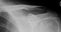
Fig.1
Initial fracture sustained at the
superiomedial angle of scapula
He had a background history of epilepsy for which he was taking
Sodium Valproate, but this was ruled out as a contributing
factor to his current injury. He was a left hand dominant
builder who was a smoker and lived with his brother.
On
examination there was diffuse fullness around the medial border
of the scapula. In elevation, he had altered scapula thoracic
rhythm; however he had a full range of active movement in both
shoulders. He was extremely tender over the superior medial
angle of the scapula and there was palpable crepitus in this
area.
 Radiographs
of the left shoulder revealed a fracture of the superomedial Radiographs
of the left shoulder revealed a fracture of the superomedial
angle of the left dominant scapula (Figure 1). He was advised to
use a sling to ease his symptoms until his shoulder felt more
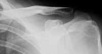
comfortable. The radiographs were repeated 8 and 12 weeks later
and the fracture was noted to be healing (Figure 2).
Fig.2
Healed scapular fracture
12 weeks later
He did not require any specialised physiotherapy regime, and
regained full function at 12 weeks post injury. He remained
asymptomatic at a 2 year follow up.
Discussion :
Scapular
fractures are relatively rare, constituting only 1% of all
fractures and 5% of those involving the shoulder, as the scapula
is well protected by overlying muscles (2). Since the scapula
does not support heavy loads, the functional repercussions are
not serious unless the glenoid is involved.
Among
scapular fractures, those of the superior portion are rare and
an avulsion fracture of the superomedial angle, as in this case
has not been described with this mechanism of injury. Due to
their rare nature, these types of fractures have not been
included in recent proposed classification of scapular fractures
(1,7).
The
most common mechanisms of scapular fractures are high-energy
motor vehicle collisions and direct violent trauma (2, 7), often
associated with other injuries. In this case however the most
likely mechanism is avulsion in the absence of violent direct
trauma. The anatomical location of the fracture in relation to
muscular attachments supports this mechanism.
Three
indirect mechanisms of scapular fractures have been described.
Uncoordinated muscle contracture due to electroconvulsive
therapy, electric shock (2, 4, 5) and more rarely epileptic
seizures (7) is one postulated mechanism. This particular
patient was epileptic, but there was no history of fitting.
Another mechanism is resisted muscle pull as a result of trauma
or unusual exertion (4). Supporting evidence lies in the fact
that this mechanism is thought to correlate with fractures of
the superior border of the scapula (4). The final mechanism is
avulsion of ligamentous attachment (4). This information
suggests that it is important to consider scapular fractures in
the differential diagnosis of shoulder pain depending on the
mechanism of injury.
If
we consider the anatomy of the muscular attachments of the
scapula in relation to this injury, the serratus anterior and
levator scapulae were the most likely culprits. Serratus
anterior is attached to the anterior surface of the medial
border of the scapula and extends to the medial potion of the
superior border. It protracts the scapula holding it against the
thoracic border as well as rotating the scapula. It has been
suggested that avulsion fractures of the inferior border of the
scapula are associated with serratus anterior (4). Serratus
anterior avulsion fracture may also be associated with winging
of the scapula due to loss of function (3, 4), which was not
noted in this case. Levator scapulae is attached to the
posterior surface of the superior part of the medial border of
the scapular. It elevates the scapula and tilts its glenoid
cavity inferiorly by rotating the scapula. We postulate that the
fracture occurred due to a strong contraction of these two
muscle groups acting on the superomedial angle of the scapula.
Despite being an avulsion injury, the fracture fragment was only
marginally displaced due to the superior fibres of the
subscapularis, which extend just onto the thoracic aspect of the
superomedial angle of the scapula. Similar avulsion fractures
have been described involving the omohyoid muscle (2, 4, 9),
however all these reports have included traumatic incidents in
the history.
This
fracture was successfully treated conservatively as illustrated
in the X-rays initially and 12 weeks later. Most scapular
fractures may be successfully treated conservatively, however
instances of non-union have been described (1, 3). These cases
occurred with fractures of the spine and body of the scapula
with the latter being partly due to ribs and muscle lodging
between bone. However this case was free of complications such
as non-union and snapping syndrome, which were also absent at a
2 year follow up.
Reference :
- Ada JR, Miller ME. Scapular Fractures: Analysis of 113
Cases. Clinical Orthopaedics and Related Research 1991; 269
174-80
- Arenas AJ, Pampliega T. An unusual kind of fracture. Acta
Orthop Belg 1993; 59(4) 398-400
- Gupta R, Sher J, Williams G et al. Non-Union of the
Scapular Body: A Case Report. J Bone and Joint Surg 1998;
80-A(3) 428-30
- Heyse-Moore GH, Stoker DJ. Avulsion fractures of the
scapula. Skeletal Radiol 1982; 9(1) 27-32
- Kotak BP, Haddo O, Iqbal M, Chissell H. Bilateral
scapular fractures after electrocution. J R Soc Med 2000; 93
143-4
- Marra G, Stover M. Glenoid and scapular body fractures.
Current Opinion in Orthopaedics 1999; 10 283-8
- McAtee SJ. Low-energy scapular body fracture: a case
report. Am J Orthop 1999; 28(8) 68-72
- Moore KL, Agur AMR. Essential
Clinical Anatomy. Baltimore:Williams & Wilkins 1995
- Williamson DM, Wilson-MacDonald J. Bilateral avulsion
fractures of the cranial the scapula. J Trauma 1988; 28(5) 713-4
|
|
This is a peer reviewed paper Please cite as
:Kevin
Kulendra: Unilateral
Superiomedial Avulsion Fracture of the Scapula - A Case
Report
J.Orthopaedics 2007;4(2)e6
URL:
http://www.jortho.org/2007/4/2/e6 |
|
|

