|
Gadegone
W M.*, Salphale YS, Sonwalkar Hemant
A, Nagtode Pankaj P, Navghare Shishir M
*Chandrapur
Multispeciality Hospital, Mul Road, Chandrapur 442401, India
Address for Correspondence
Dr.Yogesh
.S. Salphale
Shushrusha,
Opp.Z.P,Chandrapur 442401 India
Tel: (C)0091 7172 250131/263773
Fax: 0091 7172 255600
E Mail: yogeshsalphale@gmail.com
|
|
Abstract
A
variety of tumors or tumorous
conditions occur in the hand, but they are usually
benign. The hand has limited free space and exquisite
sensitivity, and even small
histologically benign masses can cause significant
swelling, pain, and disability.
Giant
cell tumor [GCT] or Osteoclastoma is a benign tumor which is
locally aggressive and has a tendency for local recurrence.
GCTs form
about 4-5% of all primary bone tumors. 80% of the patients are
above the age of 18 years and there is a distinct female
predominance.
85-90%
of the cases occur in the long bones, the sites most commonly
affected being the lower end of the femur, upper end of the
tibia, the lower end of the radius and sacrum. Only 2% of giant
cell tumors occur in the hand. We are presenting a case of a
giant cell tumor of the Proximal
phalanx of ring finger which is a very rare site for such
a tumor and presenting a follow up of 60 months following
resection and reconstruction by iliac crest graft .
J.Orthopaedics 2007;4(2)e5
Case
Report:
 A
15 year-old female presented to us with the complaints of pain
and swelling of her left ring finger since
three and half months. There was no history of trauma or any constitutional
symptoms. The swelling had gradually increased in size and there
was a gross restriction of movements of the affected ring
finger .
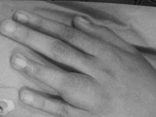
On physical examination, there was a localized swelling over the
left ring finger. The skin overlying the swelling
was free and the movements of the metacarpo phalangeal
joints were painful and restricted.(Fig 1) A
15 year-old female presented to us with the complaints of pain
and swelling of her left ring finger since
three and half months. There was no history of trauma or any constitutional
symptoms. The swelling had gradually increased in size and there
was a gross restriction of movements of the affected ring
finger .
On physical examination, there was a localized swelling over the
left ring finger. The skin overlying the swelling
was free and the movements of the metacarpo phalangeal
joints were painful and restricted.(Fig 1)
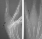
Radiographs
revealed an expansile osteolytic
lesion of proximal phalanx
of ring finger extending upto the
metacarpo phalangeal joint. (Fig 2) Extensive local bony
destruction, cortical breakthrough, and soft
tissue expansion was
noted. A fine needle aspiration cytology was done and the
diagnosis of
a giant cell tumor was confirmed.
 Grossly
the tumor consisted of brownish cheesy material which
had involved the entire phalanx. Histopathological
examination showed a well vascularized,
highly cellular tissue consisting of stromal mononuclear
cells and multinucleated giant cells present in close
association with each other.
Stromal cells were numerous, predominantly round to oval
with foci of spindling, mild degrees of atypia and occasional
mitosis. The areas of fibrous muscle tissue was seen with a few
dead bony pieces . Grossly
the tumor consisted of brownish cheesy material which
had involved the entire phalanx. Histopathological
examination showed a well vascularized,
highly cellular tissue consisting of stromal mononuclear
cells and multinucleated giant cells present in close
association with each other.
Stromal cells were numerous, predominantly round to oval
with foci of spindling, mild degrees of atypia and occasional
mitosis. The areas of fibrous muscle tissue was seen with a few
dead bony pieces .
The
tumor was carefully removed and the proximal and distal joints
inspected. Articular cartilage of the metacarpophalangeal joint
was not visible and therefore the decision of resecting the
proximal phalanx along with the articular surface of the phalanx
was carried out. The periosteal
sleeve and
the distal articular surface
of the proximal phalanx was left intact.
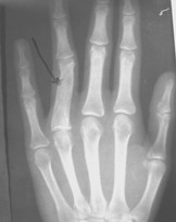 The
fibro-osseous cartilage portion of the iliac graft
was taken mimicking the phalanx in size and shape. The
graft was harvested within
the remnant of the periosteal
sleeve .Temporary fixation with K wire was done. The
K wire was removed after three months.(Fig 3) The patient
was reviewed for 3,6,9 12 months initially and thereafter every
yearly. The
fibro-osseous cartilage portion of the iliac graft
was taken mimicking the phalanx in size and shape. The
graft was harvested within
the remnant of the periosteal
sleeve .Temporary fixation with K wire was done. The
K wire was removed after three months.(Fig 3) The patient
was reviewed for 3,6,9 12 months initially and thereafter every
yearly.
The
serial xrays within showed
good incorporation and consolidation of the graft with excellent
functional results. (Fig 4)There was no signs of the diseases
elsewhere in the body
At 60
months follow-up, there was no signs of recurrence both
clinically and radiologically with good functional recovery
commensurate with her demands of daily activities. There is also
a formation of good proximal articular surface of the
phalanx.
Discussion :
Giant cell tumors
of the bones of the hand are rare accounting for only 2% of
cases and phalangeal location of the tumor is more common than metacarpals
.Giant cell tumor of the bone is a benign, but locally
aggressive lesion. It is a relatively rare tumor composed of
connective tissue stromal cells having the capacity to recruit
and interact with multinucleated giant cells that exhibit the
phenotypic features of osteoclasts
[1]. In a review of
all cases of giant cell tumors of the bone at the Mayo Clinic
over a 50 year period ending in 1994, only 13 GCTs involved the
hand, and only three involved the thumb [2] . When present in
the hand they generally extend to the articular cartilage and
are eccentrically located.[3]
GCT
of the hand seems to represent a different lesion than
conventional GCT in the rest of the skeleton. There is an 18%
incidence of multicentric foci indicating that a bone scan
should be a part of routine workup of these tumors
[4] Overall they appear in a younger age group and recur more
rapidly in the hand than they do in other locations. In a series
of 326 GCTs studied , Picci et al concluded that only six histologically proven cases had
an open epiphyseal plate which accounted for 1.8% of their
series. [5] The mean age of patients presenting with giant cell
tumors is 32, whereas the mean age for presentation with a GCT
of the hand is even less (only 22). [6]
They
also have a shorter duration of symptoms averaging six months or
less before a diagnosis is made as in our case
[7] Even
though cortical disruption takes place , the periosteum is rarely
breached [8]
Despite
the fact the GCT is not a sarcoma, the extent of tumor at the
time of diagnosis and the high recurrence rate following limited
resection often dictate the need of an enbloc resection through
normal tissues to prevent local recurrence of the lesion. Such a
treatment creates a significant skeletal defect and a
challenging reconstructive problem. The various treatment
modalities described in literature are curettage, curettage and
bone grafting, irradiation, amputation, and resection with reconstruction. [1,2]
Local
resection of the involved phalanx with autograft or allograft
replacement is the preferred surgical treatment for several
reasons. In addition curettage with or without bone grafts has
resulted in recurrence rates of about 90%. Thus curettage is an
unacceptable form of treatment. Secondly, although amputation
may prevent recurrence, it is cosmetically deforming and
decreases the function of the hand . [7 ]
The proximal
phalangeal joint reconstruction can be achieved by metatarsal
substitution, a combined iliac crest and metatarsal head graft
[9] and prosthetic replacement [10 ]. The use of radiation
as the primary treatment of a giant cell tumor of bone has been
associated with malignant transformation .
However,
in our case we used the remnant of the periosteal sleeve for
harvesting the iliac crest graft. The aim of our paper is to
demonstrate the efficacy of the iliac crest graft
and the consolidation and molding according to the
functional demands of the hand. It also depicts the
effect of mobilization of the ring finger in moulding the iliac
crest graft on the metacarpal head and also to
demonstrate the molding of the iliac crest graft similar
to phalanx. This approach allowed wide resection for
local tumour control, re-established structural integrity,
preserved metacarpophalangeal joint motion and allowed
early motion.
Reference :
-
Giant cell
tumor of bone. An analysis
of 218 cases J Bone
and Joint Surgery [Am] 1970; 52:619-64.Goldenberg RR, Campbell
CJ, Bonfiglio M.
-
Giant Cell
Tumors of the Bones of the Hand. Journal of Hand Surgery1997. 22A: 91-98.Athanasian, EA.
-
Radiology of the Hand. 254-255. 1986 Cornelis, JP
-
Giant cell tumors of the bones of hand. Journal of Hand Surgery 1980; 5:39-50. Averill RM, Smith RJ, Campbell CJ
-
Giant-cell
tumor of bone in skeletally immature patients J-Bone Joint Surg-Am. 1983 Apr; 65(4): 486-90 Picci-P,
Manfrini M, Zucchi V, GherlinzoniF, RockM, BertoniF, Neff-JR
-
Giant Cell
Tumor of the Distal Phalanx of the Hand in a Child. Clinical
Orthopedics and Related Research. 310: 200-207, 1995 Yin, Y.
-
Giant cell
tumor. A study of 195 cases. Cancer 1970; 25:1061-70. Dahlin DC,
Cupps RE, Johnson EW Jr.
-
Dahlin's bone
tumors: general aspects and data on 11,087 cases. New York, NY:
Lippincott-Raven; 1996: 463 Unni KK
-
Experimental
and clinical autogenous distal metacarpal reconstruction.
Clinical Orthopedics 1971; 74:129. Kettlekamp DB, Rampsey P.
-
Case Report of a
Giant cell tumor of the second metacarpal bone and implantation
of a cement prosthesis. Hand Chirurgie 1979; 11:251. Dingles WR, Rolle HJ
|

