|
Abstract:
The
utility and efficacy of VAC therapy has led to its use in wound
management among many specialties.
Challenges may be encountered, however, when the VAC
device is applied to irregular surfaces.
The technique presented in this case report illustrates
an efficient and effective method for the circumferential
placement of the VAC device around external orthopedic hardware
that provides a reliable seal and reduces the difficulty of
dressing changes.
J.Orthopaedics 2007;4(2)e35
Introduction:
The
use of vacuum-assisted closure (VAC) for wound management is a
widely accepted and utilized practice.
This device has been shown to decrease local edema and
bacterial loads, increase blood flow, and remove excess fluid
from the wound, promoting the formation of granulation tissue
and accelerating healing by secondary intention (1-4).
The
clinical applications for this therapy are vast and include
treatment of traumatic wounds, diabetic ulcers, venous stasis
ulcers, diabetic foot ulcers, and wounds with exposed bone and
hardware (5). In
the typical application of the VAC dressing (Kinetic Concepts,
Inc; San Antonio, TX), a foam sponge is applied to the wound,
covered with an occlusive film dressing, and uniform negative
pressure is applied (4,6).
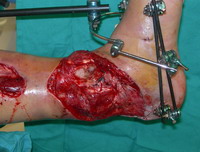
Challenges are encountered with application of the VAC
device in proximity to external orthopedic hardware (Fig. 1).
When there is inadequate skin between the wound and
fixator, the adhesive film can be applied to the hardware
itself. Wrapping
the entire frame is a solution to the problem (7), though it too
presents challenges. Tenting and tearing of the occlusive film can occur.
Moreover, the adhesive film dressing can be difficult to
remove from the fixation device and tends to leave a residue of
adhesive. We
present an efficient and effective technical adaptation for the
placement of VAC therapy around external orthopedic hardware.
|
 |
|
Figure
1: Distal
tibia and fibula fractures with soft-tissue loss after
debridement and reduction with an external fixator. |
A
54 year old male involved in a motorcycle accident presented
with a grade 3B open fracture with concurrent dislocation of his
left ankle with extensive soft tissue damage. The ankle was reduce and stabilized with a combination of
internal and external fixation techniques.
Clinical instability prevented early flap coverage of the
wound. The presence
of an external fixator in proximity to this extensive wound made
application of the VAC by conventional means quite difficult.
The technique of VAC application was adjusted to
accommodate this challenge.
As
with other techniques, the polyurethane foam is cut to the
dimension of the wound. A
non-adherent dressing is usually applied to the wound base to
facilitate sponge removal during subsequent dressing changes.
Mepilex (Mölnlycke Health Care; Norcross, GA), a
silicone backed foam dressing, can be applied to the surrounding
skin to prevent maceration of the intact skin by serving to wick
fluid from the system. The
silicone backing of the Mepilex allows it to stick to intact
skin and maintain its position without adhesive.
The foam sponge can be stapled to the Mepilex to secure
its position directly over the wound bed and prevent shifting
with suction application. This
avoids stapling the sponge to the skin in patients for whom the
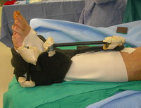
dressing change is performed without anesthesia. Prominent points of the fixator can be padded to reduce the
risk of leaking, particularly in areas that are subjected to
pressure (Fig. 2).
|
 |
|
Figure
2: Foam sponge dressing is place over the open
wounds and the surrounding skin is protected with a
non-adherent dressing. |
An impervious stockinette (Convertors, Allegiance Healthcare
Corp; El Paso, TX), used in surgical draping, is used to provide
the occlusive covering for the system.
The cloth portion of the stockinette is removed and one
of the corners is cut off of the sealed end.
The stockinette is placed over the extremity and fixator
and the toes or foot is passed through the hole created in the
end. The
stockinette is then unrolled over the frame and cut to the
appropriate length. A strip of the adhesive film is placed over the distal end of
the stockinette, sealing it to the intact skin.
The proximal end is sealed by applying traction to the
anterior aspect of the stockinette and the sides and posterior
aspect of the stockinette is sealed to the leg with occlusive
dressing. The free
edges of the stockinette are then sealed to one another and the
anterior aspect of the leg.
A slight offset of the edges near the leg facilitates
sealing. The suction interface is then applied to a part of the
stockinette that overlies the polyurethane sponge. The stockinette is then tucked into the interstices of the
external fixator and suction is applied.
In the event of tenting of the stockinette, suction can
be released and the stockinette can be repositioned.
The VAC then conforms to the leg and external fixation
device (fig. 3).
|
 |
|
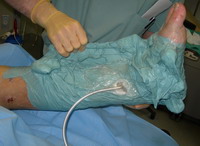
Figure
3: Completed
VAC application. The
stockinette is sealed on both the proximal and distal
edges with adhesive and conforms to the fixator. |
In the event of dressing change, removal of a VAC device applied
using this technique is quite simple. Adhesive must only
be removed from two small areas of the lower extremity, rather
than from both the external fixator and skin. This makes
for rapid dressing changes that are well tolerated by patients
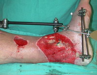
and surgeons alike. In the case presented in this report,
the wound improved remarkably with reduced edema and granulation
tissue growth. The surrounding skin was well preserved
(Fig. 4).
|
 |
|
Figure
4: Lower extremity wound after 18 days of VAC
therapy utilizing the stockinette technique |
Discussion :
With extremity wounds found in association with external
hardware, one of the main limitations to the effective use of
VAC therapy is the ability to establish and maintain an adequate
seal (8). The
technique presented here reduces this difficulty by shifting the
points that are sealed away from the hardware and wound.
The stockinette does not adhere to the fixator, which
allows greater flexibility in conforming to the contour of the
limb and hardware. This technique reduces the amount of adhesive film in contact
with the skin and external hardware, allowing for more tolerable
dressing changes and preventing the deposition of adhesive
residue on the fixator. The
extremity distal to the VAC dressing can easily be monitored for
perfusion by assessing distal pulses, capillary refill and
sensation. The
technique outlined in this paper provides a safe, effective and
efficient means of applying the VAC dressing in the setting of
challenging wounds with associated external orthopedic hardware.
Reference :
References
-
Argenta LC, Morykwas MJ. Vacuum-assisted closure: a new
method for wound control and treatment: Clinical experience.
Annals of Plastic Surgery. 1997 Jun;38(6):563-76.
-
Morykwas MJ, Argenta LC, Shelton-Brown EI, McGuirt W.
Vacuum-assisted closure: A new method for wound control and
treatment: Animal studies and basic foundation. Annals of
Plastic Surgery. 1997 Jun;38(6):553-62.
-
Argenta LC, Morykwas MJ, Marks MW, DeFranzo AJ, Molnar JA, David LR. Vacuum-Assisted
Closure: State of Clinic Art. Plastic and Reconstructive
Surgery. 2006 Jun;117(7 Suppl):127S-142S.
-
DeFranzo AJ, Argenta LC, Marks MW,
Molnar JA,
David LR, Webb LX.. The use
of vacuum-assisted closure therapy for the treatment of lower
extremity wounds with exposed bone. Plast Plastic and
Reconstructive Surgery. 2001 Oct;108(5):1184-91.
-
Geller SM, Longton JA. Ulceration of pyoderma gangrenosum
treated with negative pressure wound therapy. Journal of the
American Podiatric Medical Association. 2005
Mar-Apr;95(2):171-4.
-
Herscovici D, Sanders RW, Scaduto JM., Infante A,
DiPasquale T. Vacuum-assisted wound closure (V.A.C. therapy) for
the management of patients with high-energy soft tissue
injuries. Journal of Orthopedic Trauma. 2003
Nov-Dec;17(10):683-8.
-
Ozer K. Smith WA. Simple Technique for Applying
Vacuum-Assisted Closure Therapy Over the Circular Type External
Fixation Device. Annals of Plastic Surgery. 2006
Jun;56(6):693-4.
-
Greer SE, Duthie E, Cartolano B, Koehler KM,
Maydick-Youngberg D, Longaker MT. Technique for applying subatmospheric pressure dressing
to wounds in difficult regions of anatomy. Journal of wound, ostomy, and continence nursing. 1999
Sep;26(5):250-3.
|

