| CASE
REPORT |
|
Benign Fibrous
Histiocytoma of Sacroiliac Joint
|
|
* Korhan
Ozkan, Kerem Bilsel, Harzem Ozger, Feyza Unlu Ozkan,
Zafer Coban
*Medical Faculty of Istanbul
University, Department of Orthopedics and Traumatology, Istanbul,
Turkey
Address for Correspondence
Korhan Ozkan, MD,
Istanbul Universitesi Ortopedi ve Travmatoloji
A.B.D, ISTANBUL/TURKEY
Tel: +90 (212) 414 20 00 ( 3 2875)
Fax: +90 (216) 473 50 08
Cell Phone: +90 (532) 224 24 48
E-mail:
feyzamd@yahoo.com,
korhanozkan@hotmail.com |
|
Abstract
Benign fibrous histiocytoma of bone is an
extremely rare tumor with fibroblastic andhistiocytic
differentiation.¹ It is usually seen between third and sixth
decades of life. ² The most presenting symptom is pain. ³ The
tumor usually has a well defined radiolucent zone, with a soap
bubble appearance and sclerotic rim and no periosteal reaction.¹
Key words: sacroiliac joint, pain, histiocyte ,benign
J.Orthopaedics 2006;3(1)e9
Case Report:
 A
65 years old woman was seen after having had pain in her right
pelvis for more than a one year. The physical examination
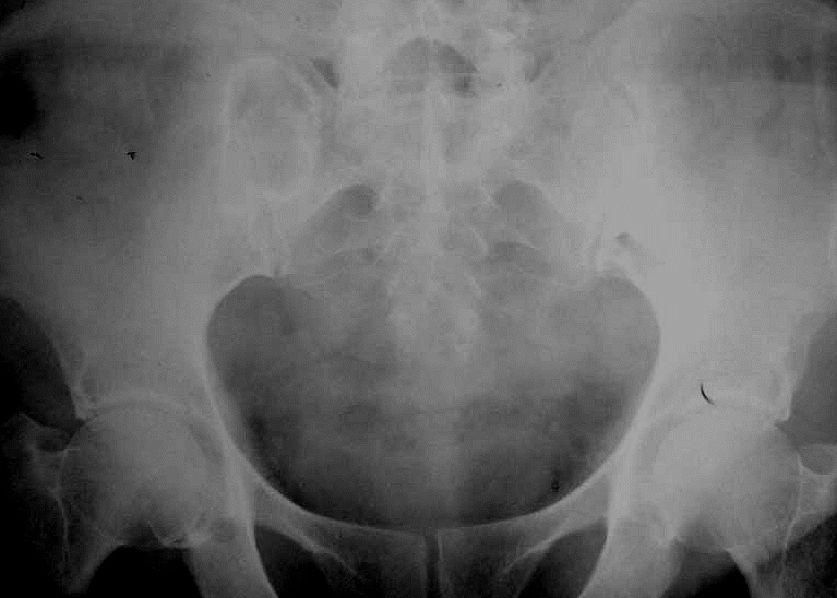
revealed no findings. Plain radiographs displayed a well
circumscribed lucent lesion with sclerotic rim in her sacroiliac
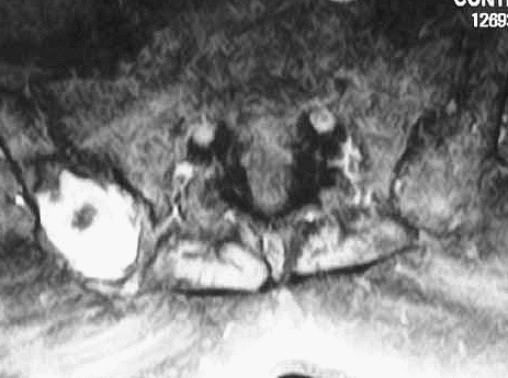
joint (figure 1). MRI displayed a well circumscribed lesion
(figure 2). An open biopsy was performed and pathological
specimens showed proliferation of fibroblasts as benign oval
spindle cells mixed with mononucleated and multinucleated
histiocytes. Macroscopic yellow zones of tumor was found to be A
65 years old woman was seen after having had pain in her right
pelvis for more than a one year. The physical examination
revealed no findings. Plain radiographs displayed a well
circumscribed lucent lesion with sclerotic rim in her sacroiliac
joint (figure 1). MRI displayed a well circumscribed lesion
(figure 2). An open biopsy was performed and pathological
specimens showed proliferation of fibroblasts as benign oval
spindle cells mixed with mononucleated and multinucleated
histiocytes. Macroscopic yellow zones of tumor was found to be
 composed
of variable amount of large round cells with vacuolated
cytoplasm. No pleomorphism and atypical mitosis was detected.
Following open biopsy one month later, intralesional curettage
followed by spongious chips greft insertion was performed.
Patients symptoms disappeared completely 1 week after operation
and started to walk with full weight bearing two weeks after.
One year later the patient was alive with no evidence of
disease. composed
of variable amount of large round cells with vacuolated
cytoplasm. No pleomorphism and atypical mitosis was detected.
Following open biopsy one month later, intralesional curettage
followed by spongious chips greft insertion was performed.
Patients symptoms disappeared completely 1 week after operation
and started to walk with full weight bearing two weeks after.
One year later the patient was alive with no evidence of
disease.
Discussion :
It is impossible to differentiate benign
fibrous histiocytoma from metaphyseal fibrous defect on the
basis of histologic and electron microscopic findings.
Metaphyseal fibrous defects are usually seen in children between
the ages of four and eight while benign fibrous histiocytoma is
seen between third and sixth decades .Metaphyseal fibrous
defects are located in long tubular bones metaphyseal region
whereas benign fibrous histiocytoma can be located in vertebrae,
pelvis, ribs other than metapyseal regions of long bones. Pain
is the major symptom of benign fibrous histiocytomas and these
lesions can be cured effectively with curettage and grafting.
Reference :
-
Clarke BE, Xipel JM, Thomas DP. Benign
fibrous histiocytoma of bone. Am J Surg Pathol 1985;9:806-15
-
Dorfman HD, Czerniak B. Bone Tumors .St
Louis, Missouri:The Mosby Company, 1998:493-513
-
Bertoni F, Calderoni P, Bacchini P. Benign
fibrous histiocytoma of bone. J Bone Joint Surg Am
1986;68:1225-30.
|
|
This is a peer reviewed paper Please cite as
: Korhan
Ozkan: Benign Fibrous
Histiocytoma of Sacroiliac Joint
J.Orthopaedics 2006;3(1)e9
URL:
http://www.jortho.org/2006/3/1/e9 |
|
|

