|
Abstract:
Lateral end clavicle fractures are uncommon but often lead to
delayed union or non-union. This is particularly the case with
displaced fractures treated non-operatively. We report our
medium term results of patients with lateral end of clavicle
fracture treated with operatively with hook plate fixation.
20 consecutive cases of lateral end of clavicle fractures were
retrospectively reviewed to assess the union rate and functional
outcome at a mean of 6.5 years post-operatively.
There were (14 male and 6 female)
patients with a mean age of 39 years. 17 (85%) patients had
acute fractures and 3 (15%) had
established non-unions at the time of surgery.
Our management consisted of hook plate fixation in all 20
patients with ancillary bone grafting in three patients with
non-union. Osseous union was achieved in all 20 patients with
surgical stabilization at mean of 9.5 weeks. All patients had
the plate removed at average 6.5 months. 23% of patients had
persistent impingement symptoms and pain post-operatively, with
9% of the patients undergoing Subacromial decompression at a
later date with resolution of the symptoms subsequently. The
average DASH score at the time of the final follow up was 9.8
(0-28) and Oxford score was 17 (12-27). At the time of final
follow up. 86% of patients had excellent to good results at
final follow up.
Hook plate fixation for lateral end of clavicle fracture is a
safe and reliable option to achieve union in this difficult
group of patients with good results over medium term with a
proportion of patients experiencing impingement symptoms.
J.Orthopaedics 2010;7(4)e4
Keywords:
Distal clavicle fracture; Hook plate; Lateral end clavicle;
impingement
Introduction:
Fractures of the clavicle are one of the most common injuries
accounting for 5% of all fractures and 44% of shoulder girdle
injuries (1,2). Lateral end of clavicle fractures account for
12-15% of these injuries. (3) There is no common consensus on
operative versus conservative treatment of these injuries.
However in displaced cases mostly surgery is performed because
of the high non-union rate often seen with these fractures.
Although there has been no prospective randomized studies
comparing different treatment modalities for these fractures,
various case series has shown non union rates as high as 22-55%
with non operative treatment in comparison to union rates of
90-95% with operative fixation (4,5).
Previous published literature has shown favourable short term
outcomes with Hook plate fixation with regard to radiological
union rates and clinical
outcomes (6,7). The aim of our study was to assess the medium
term outcome following fixation of lateral end of clavicle
fractures with hook plate.
Materials
and Methods:
Between 2000- 2004,
20 consecutive cases of lateral end of clavicle fractures were
treated in our upper limb unit with hook plate fixation. All the
patients were operated on by the senior author (JS) or under his
direct supervision. All patients with displaced Neer Type 2
clavicle fractures were included in the study. Patients with
ipsilateral upper limb fractures, polytrauma and open fractures
were excluded from the study. Standard surgical technique was
followed using the hook plate (Synthes West Chester, PA).
All patients were immobilised for a
period of 4-6 weeks with mobilisation thereafter within the
comfort limits. All patients were reviewed
retrospectively at 29.7 months and again more recently to assess
the union rate clinically and radiologically and the functional
outcome using the DASH and Oxford scores by independent
reviewers. Patient demographics, occupation, mechanism of
injury, date of injury, time to surgery, time to union,
functional outcome and return to work were documented and
analysed.
Patients were categorized into 2 groups: acute fractures treated
surgically within 4 weeks of injury and delayed (non-union)
group referred to our unit after conservative treatment
elsewhere. All the patients were followed up until clinical and
radiological union was achieved. All patients underwent planned
removal of the hook plate once the union was achieved.
Results :
There were 14 male and 6 female
patients with a mean age of 39 years (range 21-65 years).
Mechanism of injury included road traffic accident in 8 (38%),
fall in 8 (38%) and sports injury in 5 (24%) patients.
17 (85%) patients had acute
fractures and 3 (15%) had non
union (previously treated conservatively) at time of surgical
intervention. Our management consisted of hook plate fixation
in all patients with ancillary bone
grafting (DBX and Osigraft each) in three patients with
non-union. Osseous union was achieved in all 20 patients
at a mean of 9.5 weeks (range 6 12 weeks). The patients were
followed up thereafter and their scores at average follow up of
29.7 months (11-44 months) was DASH 8.4(0-28) and Oxford score
of 15 (12 27). 18 out of 20 patients returned to work at
average time of 14.1 weeks. All patients had plate removal at
average 6.5 months (range 5 months-12 months) post insertion. At
the time of that follow up, complications included rotator cuff
impingement in 5 patients, which improved to some extent in 3
patients and superficial wound infection in 1 patient requiring
antibiotic. The same group of patients were reviewed again
functionally at an average follow up of 77 months (59-89
months). The average DASH and Oxford score were respectively 9.8
(0-28) and 17 (12-27). Impingement symptoms experienced by 5
patients in the 1st follow up improved over a period
of time in 3 patients with conservative management. Remaining 2
patients underwent Subacromial decompression for persistent
impingement symptoms and pain. All the 5 patients with
impingement were more than 45 years of age. At the time of
follow up 86% of patients had excellent to good results.
|
Patient |
Age |
Sex |
Profession |
Side |
Impin-
gement |
Dash score
Final F/u |
Oxford score
Final F/u |
|
1 |
61 |
F |
Teacher |
R |
No |
3.33 |
12 |
|
2 |
47 |
M |
Postman |
R |
Yes |
11.67 |
18 |
|
3 |
32 |
M |
Radiographer |
R |
No |
1.67 |
12 |
|
4 |
34 |
F |
Secretary |
R |
No |
0 |
13 |
|
5 |
42 |
M |
Editor |
L |
No |
9.17 |
16 |
|
6 |
42 |
M |
HR Consultant |
R |
No |
5.00 |
14 |
|
7 |
30 |
M |
Cleaner |
R |
No |
1.67 |
15 |
|
8 |
65 |
M |
Security Officer |
L |
SAD |
26.67 |
22 |
|
9 |
31 |
F |
Teacher |
R |
No |
28.33 |
19 |
|
10 |
29 |
M |
Radio Assistant |
L |
No |
12.50 |
19 |
|
11 |
34 |
M |
Student |
L |
No |
5.00 |
12 |
|
12 |
26 |
M |
Desk job |
R |
No |
2.50 |
13 |
|
13 |
61 |
F |
Office job |
R |
SAD |
30.30 |
43 |
|
14 |
21 |
M |
Builder |
L |
No |
2.00 |
17 |
|
15 |
25 |
M |
Manager |
L |
No |
3.83 |
15 |
|
16 |
29 |
F |
Office job |
L |
No |
4.17 |
12 |
|
17 |
59 |
M |
Manual work |
R |
Yes |
20.00 |
17 |
|
18 |
47 |
M |
Nurse |
R |
Yes |
10.80 |
15 |
|
19 |
54 |
M |
Manual work |
L |
No |
9.90 |
20 |
|
20 |
21 |
M |
IT personnel |
R |
No |
11.20 |
17 |
SAD- Subacromian decompression
Table 1: Patient demography
and scores.
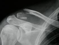
Figure
1a:Pre
op x ray case .
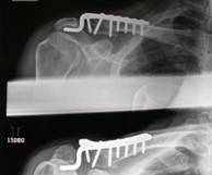
Figure 1b:
Post op x ray case 1.
Discussion :
Clavicle fractures are common injuries with the middle third
fractures accounting for 80% , medial end 5% and the lateral end
for 15% of the clavicle fractures. The non-union rates in
conservatively treated lateral end of clavicle fractures can be
as high as 22-50% (5). The delayed healing and the non-union is
associated with considerable morbidity and time off work as
shown previously by Webber and Haines (8).
The reason for the high non union rate is thought to be partly
due to the inherent unstable nature of the injury with
significant gap at the fracture site due to the attachment of
coracoclavicular ligament to the proximal fragment especially in
Neer type 2 fractures. Numerous studies have shown satisfactory
outcome with operative treatment of these fractures with
radiological union in as much as 95% of the cases (6). Various
surgical techniques have been used with good success rates and
some associated complications. Currently there is no consensus
on the ideal treatment for these injuries.
The plate design for fixation of the distal clavicle fracture
has undergone many changes. Initially called the Balser plate,
the newer design (Synthes, Switzerland) has been modified to
provide at least 2 screws to fix the lateral fragment in
addition to the hook providing additional lateral fixation and
has an oval sliding hole for dynamic compression. It also allows
the rotational movement of the clavicle during abduction and
flexion of the shoulder which reduces the incidence of implant
failure and pain, hence allowing early mobilization. Various
authors have reported satisfactory outcome with use of the hook
plate (9-11, 12, 13). The reported complications in literature
include impingement, cuff damage, acromion osteolysis, peri-prosthetic
fractures, plate migration and acromion fracture (9, 13, 14).
In our series all patients underwent planned removal of plate
once radiological and clinical union was evident at ~ 6.5 months
avoiding most of these complications. (7). Hence we recommend
removal of the plate routinely when union is achieved.
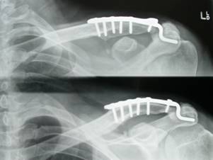
Figure 2:
Follow up x ray case 2 showing fracture healing.
Due to rarity of this fracture most of the published series has
been small numbers with a short follow up. Only one series by
Haider e al (10, 15) had mean follow up of 39 months. In our
series an initial assessment was carried out at a mean of 29.7
months to obtain short term results and subsequently we assessed
the same group of patients functionally at average of 77 months
to assess the medium term results. The average DASH score and
Oxford scores at final follow up didnt change significantly
with most of the patients (18 out of
20) returning to their pre-injury employment.
2 (10%) patients had persistent impingement even after plate
removal and failed conservative treatment and subsequently
underwent arthroscopic subacromial decompression with only
partial relief of symptoms. Both these patients were above the
age of 45 and possibly had some pre-existing risk factors for
impingement. None of the patients had
any associated acromioclavicular joint pain in short or medium
term.
Overall 86% of patients reported excellent to good results which
was no different than the short term results. This study
confirms that after the initial surgery and removal of metal
work most patients maintain a good level of function with no
further deterioration at minimum 5 years follow up with no
associated acromioclavicular joint pain. However most still have
a mild degree of discomfort related to the initial injury. Our
study was limited from the fact that it was a retrospective
analysis of a single centre series with no comparable control
group. However we do feel that it highlights the fact that
lateral end of clavicle fractures are significant injuries with
associated morbidity even at medium term despite successful
surgical treatment. The overall outcome in patients with
persistent impingement symptoms is less satisfactory despite
surgery performed in small numbers. Despite the shape of the
plate there does not seem to be any long term impact on the
acromioclavicular joint with no associated pain. This technique
is not without complication but this can be minimised by
meticulous surgical technique and timely removal of the plate.
This series highlights further the suitability of the technique
in appropriately selected patient as any one technique is not
applicable in all situations.
Reference:
-
Nordqvist A, Petersson C, Redlund-Johnell I. The natural
course of lateral clavicle fracture. 15 (11-21) year follow-up
of 110 cases. Acta Orthop Scand. 1993 Feb;64(1):87-91. PubMed
PMID: 8451958
-
Nordqvist A, Petersson C. The incidence of fractures of the
clavicle. Clin Orthop Relat Res. 1994:300:127-132.
-
Khan LA, Bradnock TJ, Scott C, Robinson CM. Fractures of the
clavicle. J Bone Joint Surg Am. 2009 Feb;91(2):447-60.
Review. PubMed PMID: 19181992.
-
Bisbinas I, Mikalef P, Gigis I, Beslikas T, Panou N,
Christoforidis I. Management of distal clavicle fractures.
Acta Orthop Belg. 2010 Apr;76(2):145-9. PubMed PMID: 20503938.
-
Rokito AS, Zuckerman JD, Shaari JM, Eisenberg DP, Cuomo F,
Gallagher MA. A comparison of nonoperative and operative
treatment of type II distal clavicle fractures. Bull Hosp Jt
Dis. 2002-2003;61(1-2):32-9. PubMed PMID: 12828377.
-
Klein SM, Badman BL, Keating CJ, Devinney DS, Frankle MA,
Mighell MA. Results of surgical treatment for unstable distal
clavicular fractures. J Shoulder Elbow Surg. 2010 Mar 23. [Epub
ahead of print] PubMed PMID: 20338788.
-
Renger RJ, Roukema GR, Reurings JC, Raams PM, Font J,
Verleisdonk EJ. The clavicle hook plate for Neer type II
lateral clavicle fractures. J Orthop Trauma. 2009
Sep;23(8):570-4. PubMed PMID: 19704272.
-
Webber MCB, Haines JF. The treatment of lateral clavicle
fractures. Injury 2003: 31(3):175-9.
-
Muramatsu K, Shigetomi M, Matsunaga T, Murata Y, Taguchi T.
Use of the AO hook-plate for treatment of unstable fractures
of the distal clavicle. Arch Orthop Trauma Surg. 2007
Apr;127(3):191-4. Epub 2007 Jan 13. PubMed PMID: 17221230.
-
Haidar SG, Singh Shergill G. Re: clavicular hook plate for
lateral end fractures: a prospective study. Injury. 2007
Feb;38(2):252-3. Epub 2006 Oct 18.PubMed PMID: 17052719.
-
Flinkkilä T, Ristiniemi J, Lakovaara M, Hyvönen P, Leppilahti
J. Hook-plate fixation of unstable lateral clavicle fractures:
a report on 63 patients. Acta Orthop. 2006 Aug;77(4):644-9.
PubMed PMID: 16929443.
-
Meda PV, Machani B, Sinopidis C, Braithwaite I, Brownson P,
Frostick SP. Clavicular hook plate for lateral end fractures:-
a prospective study. Injury. 2006 Mar;37(3):277-83. Epub 2006
Jan 23. PubMed PMID: 16430895.
-
Tambe AD, Motkur P, Qamar A, Drew S, Turner SM. Fractures of
the distal third of the clavicle treated by hook plating. Int
Orthop. 2006 Feb;30(1):7-10. Epub 2005 Oct 19. PubMed PMID:
16235083; PubMed Central PMCID PMC2254672.
-
Kashii M, Inui H, Yamamoto K. Surgical treatment of distal
clavicle fractures using the clavicular hook plate. Clin
Orthop Relat Res. 2006 Jun;447:158-64. PubMed PMID: 16505714.
-
Hackenberger J, Schmidt J, Altmann T. [The effects of hook
plates on the subacromial space--a clinical and MRT study]. Z
Orthop Ihre Grenzgeb. 2004 Sep-Oct;142(5):603-10. German.
PubMed PMID: 15472772.
|

