|
Abstract:
Objectives: Treatment of distal tibial fractures using minimally
invasive plate osteosynthesis (MIPO) technique may minimise
damage to soft tissues and the vascular integrity of bony
fragments. This is a multicentred study to assess the outcome of
patients treated with MIPO technique for distal tibial
fractures.
Methods:
A retrospective study of
27 patients from two institutions treated for distal tibial
fractures using a distal tibial locking plate through the MIPO
technique.
Results: There were 18 males and 9 females of mean age 43 years.
The mean follow-up period was 12
months (SD±4.7). According
to the AO classification system, there were 22 patients with 43A
type fractures, one 43B, two 43C and two 42A type fractures.
There were 3 open fractures as per Gustilo and Anderson
classification. Mean time to union was 4
months (SD±1.9). All patients
were fully weight bearing at 8 weeks (SD±5.0). There was one non-union in a chronic heavy smoker who
underwent autologous bone grafting at 5 months, but still failed
to unite at 9 months post operatively. In the open fracture
group; there were two delayed unions again in heavy smokers who
achieved union at 7 and 8 months postoperatively with no further
complications. There were two superficial infections treated
successfully using oral antibiotics and no failures of fixation.
There were no cases of rotational malalignment.
Conclusion: MIPO is an effective method of treatment for distal
tibial fractures. The use of indirect reduction techniques and
small incisions is technically demanding but decreases surgical
trauma to soft tissues.
J.Orthopaedics 2010;7(1)e7
Keywords:
Distal tibial fractures; locking plates; MIPO
Introduction:
Management of distal tibial fractures remains challenging1.
They are usually the result of high energy axial compression and
rotational forces. Soft tissue compromise is often severe2.
Several methods of treatment are implemented including
non-operative treatment, external fixation, intramedullary
nailing, and internal fixation with traditional implants
(standard screws and plates). However, each of these treatment
options is associated with certain challenges3.
Non-operative treatment requires prolonged immobilisation and
may be
complicated by loss of reduction and subsequent malunion4.
External fixation may lead to pin-track infections, septic
arthritis, malalignment, and delayed union5.
Intramedullary nailing
problems include the technical difficulties with distal nail
fixation, the risk of nail propagation into the ankle joint, and
the discrepancy between the diaphyseal and metaphyseal diameter
of the intramedullary canal. Open reduction and internal plate
fixation results in extensive soft tissue dissection which may
result in wound complications and infections3.
MIPO technique for distal tibial fractures offers several
theoretical advantages such as mechanically stable fracture
fixation and less disturbance of the fracture site haematoma and
the surrounding soft tissues6.
The aims of this study were to assess the outcome of patients
treated with MIPO technique for open or closed distal tibial
fractures with specific reference to fracture union, implant
failure or other surgical complications.
Patients
and Methods:
We conducted a multicentred; retrospective study of 27
consecutive patients from two institutions treated with MIPO
using the distal tibial locking plate for open or closed distal
tibial fractures and followed them up a period of 12 months
(SD±4.7). Case notes were
analysed for patients demographic parameters, follow-up reviews
and complications. Radiographs were assessed for classification
of fractures and evidence of union. Fractures were classified
according to the AO classification system. Open fractures were
graded using the Gustilo and Anderson classification7.
The operations were performed by four different surgeons.
However, all patients received cefuroxime 1.5g at induction
followed by 750mg at 8 hours and 16 hours postoperatively. Deep
vein thrombosis prophylaxis was administered as per the units
protocols. Physical
therapy was commenced first day postoperatively.
Fracture union was defined as radiological evidence of bridging
mature callus combined with clinical union as evidenced by pain
free full weight bearing. Delayed union was defined as healing
of the fracture between 5-9 months and non-union was considered
when no evidence of healing was detected after 9 months from the
operation8. Patients with a clinical rotation
difference of >15° and a clear rotation difference between both
legs as assessed on the radiographs by a senior orthopaedic
trauma surgeon were considered to have rotational malalignment.
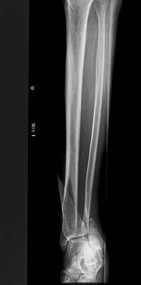
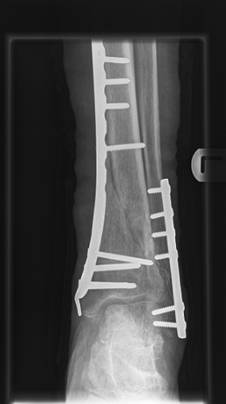
Distal tibial fracture treated with MIPO technique preoperative
and 6 months postoperative x-rays.
Results :
There were 27 patients in the study including 18 males and 9
females of mean age 43 years. According to AO classification;
there were 22 patients with 43A type fractures, one 43B, two 43C
and two 42A type fractures. The commonest cause of injury was
high-energy trauma. Twenty four patients had closed fractures
and 25 patients had closed reduction. All patients were fully
weight bearing at a period of 8 weeks
(SD ±5.0) after surgery. The mean time to union was 4
months (SD ±1.9). There was one
non-union in a 36-year-old heavy smoker who sustained a 43A type
closed fracture and was treated with the standard MIPO technique
after closed reduction of the fracture. The patient was managed
with a non weight-bearing plaster for 4 weeks then partial
weight-bearing for 6 weeks followed by full weight bearing. At
5 months postoperatively, there was no evidence of clinical or
radiological union and the patient underwent autologous bone
grafting from the iliac crest. At 9 months postoperatively,
there was still no evidence of radiological union and the
patient had pain on weight-bearing. He had normal inflammatory
markers and tissue biopsy from the fracture site was negative
for infection. There was a delayed union at 7 months in another
heavy smoker who was 46- year-old and sustained an open Gustilo
I 43A type fracture. He was treated using MIPO after closed
reduction and was allowed full weight-bearing at 11 weeks
follow-up. Another patient aged 46 years and a heavy smoker
showed delayed union at 8 months postoperatively. He had an open
Gustilo I 43A type fracture treated by wound debridement and
application of locking plate by MIPO technique. The third
patient in the open fractures group was a 27 years old patient
who presented with persistent pain around the distal end of the
scar after sustaining a Gustilo I 43C type fracture which was
treated with MIPO technique after closed reduction. He had an
area of osteolysis around the distal screw which was
subsequently removed with no further problems. There were two
superficial wound infections which were treated with oral
flucloxacillin and progressed to union. There were no failures
of fixation or implants and no rotational malalignment on
clinical and radiographic evaluation of the patients.
Discussion :
MIPO technique
has become widely practised in the operative management of
articular, metaphyseal and transitional zone fractures over the
last few years9.
It has the advantages of respecting soft tissue via small skin
incisions, minimal surgical dissection, indirect fracture
reduction and minimal hardware application. As a result, healing
time is accelerated, and complication rates are low10.
MIPO is easy to insert and gives better results with respect to
alignment correction. However, there is a learning curve and
practising the open technique initially is advisable10.
Adequate preoperative planning is mandatory, as well as accurate
surgical timing in secondary skin compromise11.
Mechanism of action depends on principles of biological
internal fixation where the aim is to produce the best
biological conditions for healing rather than absolute stability
of fixation which usually requires a fairly extensive surgical
approach to the bone. This takes advantage of indirect reduction
of the fracture and application of a bridging plate with minimal
screw insertion through stab incisions to fix the plate. The
plate functions purely as a splint rather than causing
compression of the fracture. The resulting flexible
stabilisation has been shown to give early solid union by callus
formation12. Our study supports this finding with all
patients able to fully weight bear at 8 weeks and achieving
union at an average period of 4 months after surgery. This is
consistent with results from other studies6 where
union was achieved at 22.4 and 23 weeks follow-up respectively.
Complications of nonunion and delayed union in our case series
may be associated with heavy smoking; a well recognised factor
that inhibits bone and soft tissue healing13,14.
In this study, we had two cases of
superficial wound infection, which were treated with oral
antibiotics, and no cases of deep infection or revision
fixation. These results are also similar to those previously
reported in the literature15 by Stromsoe et al who
reported stable osteo-fixation and no soft tissue complications
affecting the final result in any of his patients.
Conclusion:
MIPO technique is an effective method of treatment for fractures
of the distal tibia. The use of indirect reduction techniques
and small incisions is technically demanding but decreases
surgical trauma to soft tissues.
Reference :
-
Konrat G, Moed BR, Watson JT,
Kaneshiro S, Karges DE, Cramer KE. Intramedullary nailing of
unstable diaphyseal fractures of the tibia with distal
intraarticular involvement. J Orthop Trauma 1997;1:200-205.
-
Bourne RB. Pilon fractures of the distal tibia.
Clin Orthop 1989;240:42-46.
-
Zelle-Boris-A, Bhandari-Mohit, Espiritu-Michael, Koval-Kenneth-J,
Zlowodzki-Michael. Treatment of distal tibia fractures without
articular involvement: a systematic review of 1125 fractures.
Journal of orthopaedic trauma 2006;20(1):76-9.
-
Digby JM, Holloway GM, Webb JK. A study of function after tibial
cast bracing. Injury
1983;14:432-9.
-
Anglen JO. Early outcome of hybrid external fixation for fracture
of the distal tibia. J Orthop Trauma 1999;13:92-7.
-
Redfern DJ, Syed SU, Davies SJM. Fractures of the distal
tibia: minimally invasive plate osteosynthesis. Injury
2004;35:61520.
-
Gustilo
RB, Anderson JT. Prevention of infection in the
treatment of one thousand and twenty-five open fractures of
long bones: retrospective and prospective analyses. J Bone
Joint Surg Am 1976;58(4):453-8.
-
Bucholz RW, Heckman JD. Rockwood and
Green's Fractures in Adults. 6th ed. Philadelphia:
Lippincott, Williams & Wilkins 2006;2129-2130.
-
Pallister-Ian, Iorwerth-Awen.
Indirect reduction using a simple quadrilateral frame in the
application of distal tibial LCP-technical tips. Injury
2005;36(9):1138-42.
-
Stefano
Ghera,
Francesco Saverio Santori,
Michele
Calderaro,
Tara L
Giorgini. Minimally invasive plate osteosynthesis
in distal tibial fractures: pitfalls and surgical guidelines.
Orthopedics
2004;27(9):903-906.
-
V Pai.
A minimally invasive percutaneous plate osteosynthesis (Mippo)
for transition fractures of the distal tibia.
Journal
of Bone and Joint Surgery. (British volume)
2003;85:210.
-
Perren
SM.
Evolution of the internal fixation of long bone fractures: The
scientific basis of biological internal fixation: choosing a
new balance between stability and biology.
Journal
of Bone and Joint Surgery. (British volume)
2002;84(8):1093-2008.
-
W-Dahl A, Toksvig-Larsen S.
Cigarette smoking delays bone healing: a prospective study of
200 patients operated on by the hemicallotasis technique.
Acta Orthopaedica Scandinavica 2004;75(3):347-351.
-
Hoogendoorn J.M., Simmermacher R.K.J., Schellekens P.P.A., Van
der Werken C.
Smoking
is disadvantageous for the healing of bones and soft tissue.
Unfallchirurg 2002;105(1):76-81.
-
Stromsoe K, Eikvar K, Ovre S, Hvaal K. Miniinvasive plate
osteosynthesis of distal tibial fractures. Tidsskr Nor
Laegeforen 1999;119(29):43164318.
|

