|
Abstract:
Background:
The main purpose of this study was to assess the results of
surgical treatment of severe and stiff adolescent idiopathic
scoliosis with combined anterior-posterior approach in terms of
correction of deformity radiologically in coronal and sagittal
plane, clinically with SRS scoring and associated complications.
Materials and methods: A prospective
study of clinical and radiological outcome of 32 patients with
adolescent idiopathic scoliosis, treated surgically during
February 2006 to June 2008 with combined approach (anterior
release and posterior instrumentation) was performed.
Preoperative evaluation in the form of plain anterior, posterior
and lateral bending films and MRI spine to rule out any
congenital anomaly were performed. All of these patients had
Cobb’s angle > 60 with average being 73°± 13.4º and stiff
scoliosis. Single stage surgery was performed in all these
patients with anterior release, deformity correction and
posterior instrumentation in form of either a hybrid system with
proximal hooks and distal pedicle screws or total pedicle screw
construct. Multiple radiographic assessments on preoperative,
immediate postoperative and final postoperative radiographs and
scoring according to SRS-22 scoring system were performed. The
data was analysed using paired t test with p value <0.05 taken
as significant.
Results: The patients were followed
up for regular interval up to mean 1.5 ± 0.6 years (range 1 year
to 3 years). Coronal balance improved significantly from 80.7º
pre op to 26.7º post op. The average immediate post operative
correction achieved was 65% in coronal view and the loss of
correction over the period of 2 years was 7%. The sagittal
balance was very well taken care off with average post operative
sagittal curve being 25° in hypokyphotic spine and 35° in
hyperkyphotic spine. These were both significantly improved over
pre operative vaules.SRS scores were significantly improved post
operatively. Final fusion involved 8.6 vertebrae on an average
as compared to 9.91 levels from the pre operative radiographs.
This indicated significant number of levels preserved by the
combined approach. We had 3 superficial infection, 1 deep
infection and 2 implant removal in our series.
Conclusions: The anterior release and posterior instrumentation
is a good method of treatment of adolescent idiopathic scoliosis
with acceptable correction in coronal and sagittal alignment,
has less number of fused levels and acceptable rate of
complications.
J.Orthopaedics 2009;6(4)e6
Keywords:
adolescent scoliosis; anterior release and posterior
instrumentation; severe stiff idiopathic scoliosis
Introduction:
The posterior instrumentation has
been the mode of treatment of scoliosis since it was first
introduced by Harrington1 in 1960 in the form of
distraction rods and hooks. The development of Posterior
segmental spinal instrumentation systems1,2,3 with
third generation Cotrel- Dubousset (CD) implants which provides
multiple points of fixation to the spine and apply compression,
distraction, and rotation forces through the same rod; better
coronal plane correction and better control in the sagittal
plane could be achieved2,3. With these multiple
segmental fixation the complications associated with the
Harrington rods in form of loss of corrections, suboptimal
fixation, implant failure and lumbar kyphosis were eliminated2,3.
Even though the correction achieved with these posterior
instrumentation with pedicle screw and hook system was
satisfactory the long term results were high as it had longer
fusion level, screw breakage, increased tortional forces, crank
shaft phenomenon and flatback syndrome were commonly associated
in these patients2,3.
The anterior instrumentation first introduced by Dwyer and
supported by Zielke 4, 5, Kaneda6 and Hopf7
advocated that better correction could be achieved by placing
instrumentation in the vertebral bodies after anterior release
and discectomy. 90% of the rotational stability of the spine has
been shown to exist in the anterior two-thirds of the vertebral
body and disc, which is why an anterior release is such an
effective manoeuvre prior to fusion4. The anterior
instrumentation systems by Dwyer, Zielke, Kaneda have been used
with anterior release with or without posterior instrumentation
especially in lumbar or thoracolumbar curves.
The correction of scoliosis requires release of the tethering
structures such as rib heads, facet joints, intervertebral
discs, anterior longitudinal ligaments. This can be addressed
only with the combined anterior and posterior approaches so that
all the tethering structures can be released in order to make it
more flexible. There has been a recent interest in the posterior
only approach for scoliosis correction which relies on pedicle
screws at every level and correction obtained with rod rotation
and plastic deformation of stiff soft tissue structures. However
the posterior de-rotation manoeuvre may transmit torsional
forces to adjacent spinal segments, which can result in
decompensation, and may paradoxically increase the cosmetic
deformity in some cases and also the inability of posterior
segmental constructs to reliably provide de-rotation and restore
normal kyphosis in patients with hypokyphosis or lordosis8,9.
The use of anterior release in these cases along with posterior
instrumentation will avoid these complications and will also
decrease the number of fused levels.
In the current study, we have attempted to assess the results of
combined approach to posterior only approach in terms of the
amount of correction, SRS scores and shorter fusion length.
Materials
and Methods:
We had a total of 32 patients in our study (22
girls and 10 boys). The average age was 13.9 years (range, 10.7
to 18.2 years). 75% of the cases were in the age group of 12 to
16 years.
Inclusion criteria:
1.
Adolescent idiopathic scoliosis curves more than 60
degrees of Cobb’s angle which are stiff on side bending views
(correction < 25 deg.).
Exclusion criteria:
1.
congenital/ neuromuscular scoliosis
2.
curves less than 60 degrees
The pre operative evaluation was done in the form of plain
anteroposterior, lateral, side bending and traction radiograph
using long cassette (36”). The coronal measurement was taken
with the help of Cobb’s method10,11 and the sagittal
measurement was taken with the help of sagittal vertebral axis
which is a plumb line drawn from the centre of C7 vertebrae
body. Flexibility of the curves was measured by side bending
radiographs. MRI spine taken in all these patients was to rule
out any congenital anomaly.
The Lenke’s classification system for AIS was used in our study
12. According to Lenke’s classification the maximum
numbers of the cases in our study were of type I (15.6%) and V
(28.1%) (Table 1).
|
LENKE TYPE |
NO. OF CASES |
PERCENTAGE |
|
I |
5 |
15.6 % |
|
II |
3 |
9.3 % |
|
III |
10 |
10 % |
|
IV |
3 |
9.3 % |
|
V |
9 |
28.1 % |
|
VI |
2 |
6.2 % |
Table 1: Curve distribution
The single stage surgery was performed in these patients by one
surgeon. Anterior release was done either by open thoracotomy or
retroperitoneal approach in lateral decubitus position. Anterior
release included diskectomies and release of anterior
longitudinal ligament and was followed by posterior
instrumentation. Posterior segmental instrumentation with hybrid
fixation using hooks in proximal segments and pedicle screws in
distal segments was used in 22 patients while in 10 patients
total pedicle screw construct was used. The choice of hooks or
screws was made based on surgeon’s preference and size of the
pedicle. Pedicle screws were placed parallel to superior end
plates under the vision of image intensifier using specific
anatomical landmarks and various confirmatory tests used to
ensure intraosseus placement. Drill holes were undertapped by
1mm for better hold. The posterior facet fusion was done after
decorticating the laminae, posterior elements and the facet
joints.
The radiographic evaluation was done preoperatively, immediate
postoperatively and at the time of last follow up. In pre
operative radiographs we counted the levels of vertebrae fused
till the lowest stable vertebrae as recommended by the posterior
only fusion techniques13 . In post operative
radiographs we counted the number of levels actually released
and fused to achieve maximum correction. This difference gave us
the number of motion segments preserved by this technique as
compared to posterior only approach. Immediate post operative
radiographs were compared with the preoperative radiographs to
measure the correction achieved intraoperatively. Final
radiographs taken at the end of 2 years were compared with
postoperative radiographs to measure the loss of correction
which is due to dynamic nature of the curve and settling of the
implant.
The Scoliosis Research Society (SRS-22) questionnaire was used
as a quality-of-life instrument to assess patient outcomes after
operative treatment of adolescent idiopathic scoliosis. The SRS
score has 4 elements such as function, pain, self image and
mental satisfaction. All the patients were evaluated with SRS
scoring preoperatively, immediate post operatively and at
regular interval at the time of follow up in out patient
department.
Statistical analysis was done using paired t test with p value
of <0.05 taken as significant.
Results :
The
majority of our cases were of high grade (60-80degree) with the
average being 73°. Female patients dominated our study
consisting 65% of total cases. Results of curve correction are
given in Table 2. The post op correction achieved in our study
was 65% in coronal view measured by Cobb’s method which is
better than 47.5% as quoted in similar studies14,15,16.
In addition, the sagittal balance could be well achieved as post
op curve measured 25° in hypokyphotic spine and 35° in
hyperkyphotic spine. There was significant correction achieved
for all these angles as compared to the pre operative values (p
<0.01). comparision of angular deformities achieved at immediate
post surgery to the final follow up showed only a loss of
correction in coronal alignment of 1.8º [7%] while there was no
change in the sagittal balance.
|
Angular deformity |
Pre op
mean |
Immediate Post op
mean |
P
value |
Final
follow up |
|
Coronal |
80.7° |
26.7° |
<0.01 |
28.5º |
|
Sagital (Hypokyphosis) |
18° |
25° |
<0.01 |
25º |
|
Sagital (Hyperkyphosis) |
60° |
35° |
<0.01 |
35º |
p value is for paired t test.
Table 2: Table showing correction of angular deformities
on pre operative and final post operative radiographs.
When number of fused levels were compared with number of levels
expected to be fused on pre- operative radiographs (using
posterior only approach), we found that significantly less
number of vertebral levels required fusion when combined
approach was used (Table 3)
|
|
Expected fused segments* |
Actual fused segments** |
p value∞ |
|
Mean |
9.91 |
8.66 |
<0.0001 |
|
SD |
1.44 |
0.9 |
|
Table 3: comparison between pre operative and post
operative levels fused
* Expected segments fused in derived from pre operative
radiograph with respect to posterior only fusion
**actual segments fused are seen in post operative radiograph
using a combined approach.
∞ p value is for paired t test.
Changes in SRS 22 Score in the follow up period:- Self-image was
significantly improved at 3 months and maintained improvement
through 24 months. Function was significantly decreased at 3
months, but returned to baseline by 6 months. Pain was
significantly worse at 3 months, but was significantly less at
6, 12, and 24 months when compared to 3 months.
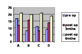
A- Pain
B- Self
image
C- Function
D-Mental satisfaction
The pre operative and post operative SRS 22 parameters were
measured pre operatively and post operatively and all were found
to be significantly improved (Table 4).
|
Variables |
Pre op
mean± SD |
Post op
mean± SD |
p - value |
|
Pain |
3.2 ± 1.09 |
4.08 ± 0.36 |
<0.0001 |
|
Self image |
1.8 ± 1.06 |
4.24 ± 0.35 |
<0.0001 |
|
Function |
2.2 ± 0.58 |
4.16 ± 0.31 |
<0.0001 |
|
Mental health |
1.8 ± 0.5 |
4.14 ± 0.42 |
<0.0001 |
p value is for paired t test
Table 4: Comparison between different SRS 22 parameters
pre operatively and post operatively.
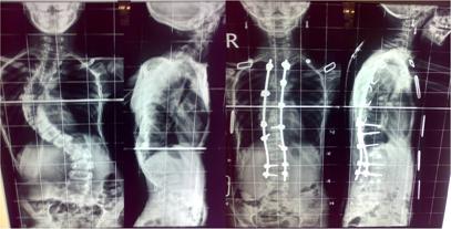
Illustrative Case Studies:
1. Master AYS 16/m: Thoraco lumbar scoliosis with right sided
major curve where anterior release from D6 to D12 and post
instrumentation moss Miami from D3 to Dl2.
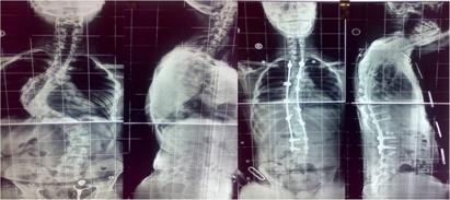
2. Miss AR 13/f: Thoraco lumbar scoliosis with left sided major
curve-post moss Miami instrumentation from D-2 to D-l2.
Complications: In our study we had 3 cases with
superficial infection which was treated with antibiotic while 1
case with deep infection was debrided in Operation theatre.
Implant was removed in this case and infection control was
achieved with repeated debridements. This patient had loss of
correction with poor results. Hook pullout was present in 1
case and 1 patient had screw breakage. Both these cases had
implants removed after bony fusion consolidated. Both these
patient had fair results. None of the cases had pseudoarthrosis,
neurological complication or death.
|
COMPLICATIONS |
NO.OF CASES |
PERCENTAGE |
|
Superficial Infection |
3 |
9.3 % |
|
Deep infection |
1 |
3.12 % |
|
Pseudoarthrosis |
0 |
0 % |
|
Crank shaft phenomenon |
0 |
0 % |
|
Thoracolumbar kyphosis |
0 |
0 % |
|
Neurological |
0 |
0 % |
|
Implant failure |
2 |
6.25 % |
Table 5: Complications
Discussion :
The goals of the scoliosis surgery include adequate and safe
correction of deformity, bony fusion to prevent further
deformity and to preserve motion segments while achieving the
above two goals. Patient satisfaction is one of the major
factors that justify these extensive surgeries. Present study
tries to analyse the results combined anterior release with
posterior fusion with respect to the goals achieved and also the
patient satisfaction in adolescent idiopathic scoliosis.
In a review of literature, Lenke et al.14 have
studied the amount of correction obtained in either anterior or
posterior fusion. An overall correction rate of 58% was achieved
in the anterior group, whereas only a 38% correction rate was
observed in the posterior group. In anterior-group patients
better spontaneous correction of the lumbar curve was
demonstrated than in posterior group patients (56% and 37%,
respectively). Good correction rates by anterior surgeries is
also reported by other authors17,18.19. In our study
too the average immediate post operative coronal correction
achieved was 65%. Over the follow up of 2 years the loss of
correction was 7%. The result of scoliosis correction in our
study was 57% as compared to preoperative measurements. The
achieved correction is very well comparable with the
contemporary literature. The sagittal imbalance was well
restored within the normal range. The average correction in
patients with hyperkyphotic spine was 35° while in hypokyphotic
spine was 25° to create a balanced sagittal curve. Creating a
stable spinal construct with sound fusion was the goal of
surgery, which was achieved in 100% (32 cases). With the help of
anterior release the spinal column was made more flexible and
amenable to posterior correction.
Recent studies by Lenke15,16 however supported
isolated posterior approach as compared to his previous studies.
He advocated that the same correction can be achieved by
isolated posterior approach compared to anterior if pedicle
screws are used rather than the hooks and at all levels rather
than at apices and end vertebrae. This observation is also
noted by Geck20, Lenke15,16, Wang21,
DiSilvestre22 , Pateder23,Hee2.
Although the correction achieved was similar with posterior only
approach using pedicle screws, the combined approach has better
correction and less number of fused levels. This has been
reported by numerous studies like Li 24,25 ,Hempfing26
, Rauzzino27.
The average number of vertebrae fused in our study was 8.6 while
calculation on the pre operative radiograph indicated fusion of
9.91 indicating preservation of at least 1.3 segment per case.
The anterior release done in these patients prior to posterior
fusion and instrumentation saved these motion segments in the
lumbar spine which resulted in shorter fused spine. This helped
us to preserve more lumbar motion segments and prevent
accelerated degeneration of the lumbar discs and the final
height achieved by the patient with less possibility of disc
degeneration and back pain in future28. Also, there
is a general concern about possible neurological injury during
the pedicle screw insertion although the incidence is quite low
and involves a definite learning curve. There is also a
commercial element affecting all posterior implants surgery
since more number of screws is required.
Complications nevertheless were present in our study in the form
of superficial infection in 3 cases and deep infection in 1 case
and 2 cases of implant failure but no incidence of neurological
complications. The cases with superficial infection healed with
one debridement and antibiotics. The case with deep infection
required debridement with implant removal and had a poor result.
Both patients with implant breakage reported after the fusion of
the segments was achieved and only implant removal was done in
these cases. A long term follow up of these patients is
essential.
The SRS 22 has been validated as a potent tool for patient
satisfaction after scoliosis surgery29 and most
important among the parameters is the improvement of self image
of the patient. In our study the self image and the mental
health are the two factors that improved very significantly as
compared to the pre operative values. Thus we have achieved
comparable results to contemporary literature with special
emphasis on better correction and saving of distal mobile
levels. According to our study the anterior release and
posterior instrumentation is a safe and effective method of
correction in adolescent idiopathic scoliosis which carries less
chance of neurological complications, better safety, high fusion
rate, lesser implant failure and manageable complication rate.
One of the limitations of the study is short follow up. Since we
have presented our early results and will be following the
cohort prospectively, we shall be presenting the mid term follow
up of these patients to assess implant failure, loss of
correction, mechanical back pain, adjacent level degeneration
and other related complications. A second limitation is relative
heterogeneity of the sample with respect to Lenke’s
classification and small sample size. An adequately powered
randomized controlled longitudinal trial will be needed to
emphasize the results of combined anterior and posterior
approach versus posterior alone surgeries in adolescent
idiopathic scoliosis.
Conclusions:
We conclude that the combined anterior and posterior method of
scoliosis correction is an effective method of correction of
scoliosis surgery in adolescent idiopathic curves in terms of
the number of fused levels and the amount of correction. The
anterior release certainly makes the idiopathic curve more
flexible and more amenable to posterior correction using
instrumentation.
Reference :
-
Irwin WD, Dixon JH, Harrington PR. Surgical instrumentation
for the management of scoliosis. J Bone Joint Surg; 1960;42:
1448. 6.
-
Cotrel Y, Dubousset J,
Guillaumat M: New universal instrumentation and spinal
surgery,
Clin Orthop Relat Res1988;227:10.
-
Farcy JP, Weidenbaum M, Michelsen CB, Hoeltzel DA, Athanasiou
KA. A comparative biomechanical study of spinal fixation using
Cotrel-Dubousset instrumentation. Spine. 1987;12(9):877-81.
-
Halm HF, Liljenqvist U, Niemeyer T, Chan DP, Zielke K,
Winkelmann W. Halm-Zielke instrumentation for primary stable
anterior scoliosis surgery: operative technique and 2-year
results in ten consecutive adolescent idiopathic scoliosis
patients within a prospective clinical trial. Eur Spine J.
1998;7(5):429-34.
-
Lowe TG, Peters JD: Anterior spinal fusion with Zielke
instrumentation for idiopathic scoliosis: a frontal and a
sagittal curve analysis in 36 patients. Spine 1993:423—426.
-
Kaneda K, Shono Y, Satoh S, et al. Anterior correction of
thoracic scoliosis with Kaneda anterior spinal system: A
preliminary report. Spine 1997;22:1358–68.
-
Hopf CG, Eysel P, Dubousset J. Operative treatment of
scoliosis with Cotrel Dubousset-Hopf instrumentation. New
anterior spinal device. Spine 1997;22:618–28.
-
Dwyer AF,
O'Brian JP, Seal PP, Hsu L, Yau AC, Hodgson AR. The late
complications after the Dwyer anterior spinal instrumentation
for scoliosis. J Bone Joint Surg 1977; 59b (1): 117.
-
Terek RM, Wehner J, Lubicky JP. Crankshaft phenomenon in
congenital scoliosis:a preliminary report. J Pediatr Orthop.
1991 Jul-Aug;11(4):527-32.
-
Cobb JR:
Outline for the study of scoliosis in instructional course
lectures. In The American Academy of Orthopaedic Surgeons:
Instructional course lectures, vol 5, Ann Arbor, Mich, 1948,
JW Edwards
-
Nash C, Moe J. A study
of vertebral rotation. J Bone Joint Surg 1969;51A:223.
-
Lenke LG, Betz RR, Haher TR, Lapp MA, Merola AA, Harms J,
Shufflebarger HL. Multisurgeon assessment of surgical
decision-making in adolescent idiopathicscoliosis: curve
classification, operative approach, and fusion levels. Spine;
2001 Nov 1;26(21):2347-53.
-
Lowe TG, Betz R, Lenke L, Clements D, Harms J, Newton P, Haher
T, Merola A,Wenger D. Anterior single-rod instrumentation of
the thoracic and lumbar spine:saving levels. Spine (Phila Pa
1976). 2003 Oct 15;28(20):S208-16.
-
Lenke, LG, Bridwell, KH, Baldus, C, Blanke, K, Schoenecker,
PL. Cotrel-Dubousset instrumentation for adolescent idiopathic
scoliosis. J Bone Joint Surg Am 1992 74: 1056-1067
-
Potter BK, Kuklo TR, Lenke LG. Radiographic outcomes of
anterior spinal fusion versus posterior spinal fusion with
thoracic pedicle screws for treatment of Lenke Type I
adolescent idiopathic scoliosis curves. Spine; 2005 Aug
15;30(16):1859-66..
-
Luhmann SJ, Lenke LG, Kim YJ, Bridwell KH, Schootman M.
Thoracic adolescent idiopathic scoliosis curves between 70
degrees and 100 degrees: is anteriorrelease necessary? Spine (Phila
Pa 1976). 2005 Sep 15;30(18):2061-7.
-
Lowe TG, Betz R, Lenke L, Clements D, Harms J, Newton P, Haher
T, Merola A,Wenger D. Anterior single-rod instrumentation of
the thoracic and lumbar spine:saving levels. Spine (Phila Pa
1976). 2003 Oct 15;28(20):S208-16.
-
Sweet FA, Lenke LG, Bridwell KH, Blanke KM. Maintaining lumbar
lordosis with anterior single solid-rod instrumentation in
thoracolumbar and lumbar adolescent idiopathic scoliosis.
Spine (Phila Pa 1976). 1999 Aug 15;24(16):1655-62.
-
Gopinathan P : Short Segment Anterior Correction Of Thoracic
Scoliosis With Single Solid Rigid Rods.( In Adolescent
Idiopathic Scoliosis). J.Orthopaedics 2008;5(2)e11
-
Geck MJ, Rinella A, Hawthorne D, Macagno A, Koester L, Sides
B, et al. Comparison of surgical treatment in Lenke 5C
adolescent idiopathic scoliosis: anterior dual rod versus
posterior pedicle fixation surgery: a comparison of two
practices. Spine (Phila Pa 1976). 2009 Aug 15;34(18):1942-51.
-
Wang Y, Fei Q, Qiu G, Lee CI, Shen J, Zhang J, Zhao H, Zhao Y, Wang
H, Yuan S. Anterior spinal fusion versus posterior spinal
fusion for moderate lumbar/thoracolumbar adolescent idiopathic
scoliosis: a prospective study. Spine; 2008 Sep
15;33(20):2166-72.
-
Di Silvestre M, Bakaloudis G, Lolli F, Vommaro F, Martikos K,
Parisini P. Posterior fusion only for thoracic adolescent
idiopathic scoliosis of more than 80 degrees: pedicle screws
versus hybrid instrumentation. Eur Spine J.
2008Oct;17(10):1336-49.
-
Pateder DB, Kebaish KM, Cascio BM, Neubaeur P, Matusz DM,
Kostuik JP. Posterior only versus combined anterior and
posterior approaches to lumbar scoliosis in adults: a
radiographic analysis. Spine (Phila Pa 1976). 2007 Jun
15;32(14):1551-4.
-
Li M, Ni J, Fang X, Liu H, Zhu X, He S, Gu S, Wang X.
Comparison of selective anterior versus posterior screw
instrumentation in Lenke5C adolescent idiopathic scoliosis.
Spine (Phila Pa 1976). 2009 May 15;34(11):1162-6.
-
Li M, Ni J, Li Y, Fang X, Gu S, Zhang Z, Zhu X. Single-staged
anterior and
posterior spinal fusion: a safe and effective alternative for
severe and rigid
adolescent idiopathic scoliosis in China. J Paediatr Child
Health. 2009May;45(5):246-53.
-
Hempfing A, Ferraris L, Koller H, Rump J, Metz-Stavenhagen P. Is
anterior release effective to increase flexibility in
idiopathic thoracic scoliosis? Assessment by traction films.
Eur Spine J. 2007 Apr;16(4):515-20.
-
Rauzzino MJ, Shaffrey CI, Wagner J, Nockels R, Abel M.
Surgical approaches for
the management of idiopathic thoracic scoliosis and the
indications for combined anterior-posterior technique.
Neurosurg Focus. 1999 May 15;6(5)
-
Ginsburg HH, Goldstein L, Haake PW. Longitudinal study of back
pain in postoperative idiopathic scoliosis: Long-term
follow-up. Presented at: the 30th Annual Meeting of
the Scoliosis Research Society; 1995; Ashville, North
Carolina.
-
Burton DC, Glattes RC. Measuring outcomes in spinal deformity.
Neurosurg Clin N Am. 2007 Apr;18(2):403-5.
|

