|
Abstract:
Introduction:
Fractures of the Calcaneum are still considered difficult to
treat with opinions varying between conservative and operative.
Method:
We had 51 cases in 45 patients of fracture Calcaneum treated by
operative as well as conservative methods. Duration of follow up
ranged from 1 year to 10.5 years, average being a 3 years follow
up.
Result:
Fractures were classified as extra—articular or intra—articular
and clinical results were evaluated according to the “Maryland
Foot Score” based on pain, gait, cosmesis and subtalar joint
motion.
Conclusion:
It was found that the clinical outcome of extra—articular
fractures was good, whatever be the method of treatment. But,
the results of intra—articular displaced fractures were better
with operative intervention than with conservative management.
J.Orthopaedics 2009;6(1)e3
Keywords:
fracture calcaneum; intraarticular; extraarticular; surgery; outcome
Introduction:
There
remains a great deal of controversy regarding the management of
calcaneal fractures 1,2: operative versus
non-operative. In operative, this controversy is further fueled
by the disagreement on which operative approach is preferable:
medial or lateral.
Treatment of calcaneum fractures has varied from Bohler’s
compression clamp 3,4 and plaster immobilization to
Essex Lopresti’s 5 percutaneous leverage techniques
and open reduction internal fixation (ORIF 6,7) by
medial 8 or lateral 9 approach or even
primary subtalar arthodesis 10.
While initially, the results of surgery in calcaneal fractures
were considered dismal, recent papers have shown good functional
outcomes after operative intervention 11,12.
The purpose of this study was to assess the results of
operative and conservative management and to attempt to decide
the best treatment protocol.
Material
Methods:
This
study included 51 cases in 45 patients with fracture (#)
calcaneum treated by conservative as well as operative methods.
Basic investigations were conventional radiographs 13
including Lateral view of hindfoot, Axial view of heel, A—P
view of foot, Broden’s view and Anthonsen’s oblique view 14.
Fractures were divided into 2 groups based on radiographs:
(a)
Extra—articular (Anterior process #/ Tuberosity #/
Sustentaculum #/ Body #)
(b)
Intra—articular (Essex – Lopresti Classification)
(Tongue type #/ Joint Depression type #)
We treated all displaced fractures operatively unless patients
refused for or were unfit for surgery.
Patients were followed up from a period
ranging from 1 year to 10.5 years, average being a 3-years
follow up. Evaluation at follow up was both clinical and
radiological (Fig 1-4).
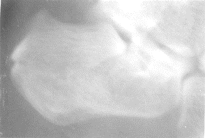 |
| Fig
1. Case 1 Pre op X-Ray |
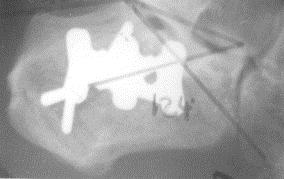 |
| Fig
2. Case 1 Post op X-Ray |
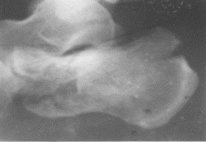 |
| Fig
3. Case 2 Pre op X-Ray |
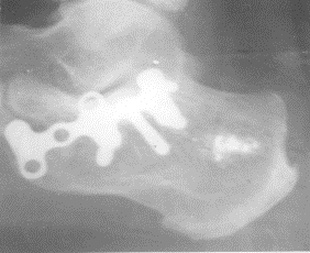 |
| Fig
4. Case 2 Post op X-Ray |
Movements at subtalar joint were measured using Mc Master’s
method15. Clinical outcome was assessed based on the
“Maryland Foot Score 16” (based on pain, gait,
cosmesis and subtalar joint motion). On a scale of 0—100,
clinical outcome was graded as Excellent (90—100), Good
(75—89), Fair (50—74) and Poor (less than 50).
The collected data was analyzed and compared with the
reported series of Robert Soeur and Robert Remy 9; Eugene Lance
and Edward Carey 17.
Results:
Of the 45 patients (37males and 8 females) included, 39 had
unilateral fracture (21 right side and 18 left side), while 6
had bilateral fractures. Most of the patients belonged to 3rd,
4th and 5th decades, youngest being 17years and oldest 70years.
Fall from height was the most common mode of injury (39
patients) while 6 patients had history of road traffic
accidents.
8 of the patients had associated injuries (2 spine injuries, 1
pelvic injury, 1 blunt abdominal trauma, 1 nasal bone fracture
and 3 had other limb injuries).
The 51 fractures included 8 extra—articular (2 anterior
process #, 2 tuberosity # and 4 body #) and 43 intra—articular
fractures (20 tongue type and 23 joint depression type #).
Out of these 51 fractures, 23 were treated conservatively and 28
were operated (Table I). 26 intra—articular and 2 displaced
extra—articular fractures were operated.
|
|
Our
Series
|
Lance
and Carey 5
|
|
Conservative
|
Operative
|
Conservative
|
Operative
|
|
(a)Treatment
given
Below
Knee Cast
Steinmann pin+Cast
Essex Lopresti
ORIF
|
15
3
5
0
|
0
0
0
28
|
104
0
27
0
|
0
0
0
17
|
|
(b) Average
duration of plaster immobilization
|
10.2
weeks
|
4.7
weeks
|
8
weeks
|
14
weeks
|
|
(c) Average
time after which full weight bearing allowed
|
14.7
weeks
|
17.4
weeks
|
12
weeks
|
29weeks
|
Table
I . Treatment Given
Based on the “Maryland Foot Score”, 75% of the operated
patients had good or excellent results while 60.8% of
conservatively treated patients fell in the same category (Table
II).
|
|
Conservative
|
Operative
|
|
Total
|
Percentage
|
Total
|
Percentage
|
|
Excellent
Good
Fair
Poor
|
7/23
7/23
7/23
2/23
|
30.4
30.4
30.4
8.8
|
8/28
13/28
4/28
3/28
|
28.6
46.4
14.3
10.7
|
Table
II. Results (Based on
MARYLAND FOOT SCORE)
We found that none of the extra—articular fractures had
fair or poor results, while a significant majority of patients
with intra—articular fractures treated with Steinmann (ST) pin
+ Below Knee (BK) Cast/ Essex-Lopresti percutaneous leverage
technique and only K—wire fixation showed fair or poor
results.
Finally Table III compares the results of BK Cast with
ORIF by Staple/Plating in Intra—articular fractures. All the
cases treated by ORIF were displaced fractures. Out of these 26
cases, 21 (80.7%) had excellent or good results. While the 4
undisplaced intra—articular fractures treated with BK Cast had excellent or good results, all the 3 displaced cases
had a fair outcome, none of them falling in the excellent or
good category indicating the need for anatomical reduction and
stable internal fixation in such cases.
|
|
INTRA—ARTICULAR FRACTURES
|
|
UNDISPLACED
|
DISPLACED
|
|
Excellent
|
Good
|
Fair
|
Poor
|
Excellent
|
Good
|
Fair
|
Poor
|
|
Below
Knee Cast
(7
cases)
|
2
|
2
|
—
|
—
|
—
|
—
|
3
|
—
|
|
Staple
or
PlateFixation
(26
cases)
|
—
|
—
|
—
|
—
|
8
|
13
|
3
|
2
|
Table
III. Comparing Below Knee Cast and ORIF by Staple or Plate in
Intra-articular fractures
Discussion:
As
in the series of Robert Soeur and Remy 9, the commonest mode of
injury was a fall from height and males sustain this fracture
much more commonly than females, most probably due to their more
active outdoor lifestyle.
We preferred operative intervention in intra articular calcaneal
fractures, except in undisplaced fractures or in patients who
refused for or were unfit for surgery, in which case,
conservative regime was undertaken.
While in the series of Lance and Carey 17, operated patients
were given post operative plaster immobilization for an average
14 weeks, we allowed earlier ankle and subtalar joint
mobilization in patients with secure internal fixation (average
4.7 weeks and in some cases even immediate post—operative)
although operated patients were generally kept non—weight
bearing
longer than those treated conservatively because internal
fixation was done in cases of displaced intra—articular
fractures. From our short experience, we sincerely believe that
early non—weight bearing joint mobilization had better results
than longer periods of immobilization.
We found the lateral approach quite satisfactory, provided a
full thickness lateral flap was raised. The problem of flap
necrosis was encountered in just 1 out of the 28 operated
patients.
The major complication in intra—articular fractures was
subtalar arthritis, which seems almost inevitable; only the
severity can be reduced by good reduction and alignment. With
time, however, the complaints of most patients reduced, the most
plausible explanation being modification of daily activities by
the patient.
Conclusion:
After
analyzing the results of both conservative and operative methods
of treatment, the following conclusions can be drawn:
1. Accurate understanding of fracture
pathoanatomy 18 and joint biomechanics 19 is an essential
prerequisite for deciding the line of management.
2. Good conventional roentgenography, which includes
lateral, axial, and if required, oblique x—rays of the
calcaneum is a must in understanding the displacement of major
fracture fragments.
3. Results of extra—articular fractures are good, whatever be
the mode of treatment.
4. Outcome of intra—articular displaced fractures is better
with operative intervention than with conservative management.
Secure fixation and early subtalar joint mobilization must be
stressed upon.
As a parting comment, it may be said that “Fractures of the Os
Calcis remain atleast partially unsolved” and the question
still lingers on—“Can we put the Humpty—Dumpty together
again?”
Reference :
-
Dror
Paley and Hamilton Hall. Calcaneal fracture
controversies.OCNA Vol. 20, No. 4, Oct. 1989. 665—677.
-
John
Randle, Hans Kreder. Should Calcaneal fractures be
treated
Surgically. CORR No. 377, Aug 2000.
-
Alexander
P. Aitken. Fractures of the OS Calcis—Treatment by closed
reduction. CORR No. 30, 1963.
-
Lorenz
Bohler. Treatment of fractures, 4th Edn,
1935.
-
Essex—Lopresti.
Mechanism, Reduction technique and results in fractures of Os Calcis. CORR No. 290, May 1993.
-
Roy
Sanders, Paul
Gregory. Operative treatment of Intra—Articular
fractures of the Calcaneus. OCNA Vol.26,April 1995. 203—214.
-
Stephen
Benirschke, Bruce Sangeorzan. Extensive intra—
articular fractures of the foot: Surgical management
of Calcaneal Fractures.
CORR No.292, July 1993. 128—134.
-
Billie
D. Burdeaux. Medial Approach for Calcaneal fractures.
CORR No. 290, May 1993. 96—107.
-
Robert Soeur, Robert
Remy. Fractures of calcaneum with
displacement of the thalamic portion. JBJS Vol.57—B,
Nov.1975. 413—421.
-
R.I.Harris. Fractures of Os Calcis –Treatment by early subtalar
Arthrodesis. CORR No.30, 1963. 100—110.
-
Paul Juliano, Hoan Vu Nguyen.
Fractures of the Calcaneus.
OCNA Vol. 32, Jan 2001.
-
Roy
Sanders. Current concepts review—displaced intra—articular fractures of the
Calcaneus. JBJS Vol. 82—A, Feb 2000.
-
Kenneth Koval, Roy Sanders. Radiological Evaluation of Calcaneal
fractures. CORR No. 290, May 1993. 41—46.
-
R.Watson Jones. Fractures and other bone and joint injuries, 1941.
-
Michael
McMaster. Technique of measuring subtalar joint movement.
JBJS Vol. 58—B, Feb 1976.
-
Roy
Sanders, Paul Fortin, Thomas Dipasquale, Arthur Walling. Operative treatment in 120 displaced intra—articular calcaneal fractures. CORR No. 290, May 1993. 87—95.
-
Eugene M.Lance, Edward J.Carey, Preston A.Wade.
Fracturesof the Os
Calcis—Treatment by early mobilization. CORR No.30,1963.
-
James B. Carr. Mechanism and pathoanatomy of intra—articular
calcaneal fractures. CORR No. 290, May 1993. 36—40.
-
Shahan
K. Sarrafian. Biomechanics of the subtalar joint complex. CORR No. 290, May 1993.
|

