|
Abstract:
Introduction
Conservative treatment of forearm fractures is fraught with complications
of cast, compartment syndrome, malunion and bayonet apposition.
Prolonged hospitalization and high cost associated with plate
fixation makes choice of treatment difficult. We prospectively
evaluated outcome and cost effectiveness of closed nailing of
forearm bone fractures in search of a better treatment.
Materials
and Methods
26 children between age group of 5 to 15 yrs with forearm fractures were
prospectively treated with closed intramedullary nailing with
Talwalkar’s Square nails during June 2006 to June 2007.
Diaphyseal fractures of both bones forearm were nailed with
flexible square nails and were given splints for 4 weeks.
Patients underwent implant removal once there was clinical and
radiological union of fractures.
Results
Of the 26
children with both bone fracture, 17 were operated within 24
hours from injury. Closed nailing was done in 18 children; mini
open ulna was done in 1 child, mini open radius in 3 children
and mini open of both radius and ulna in 4 children. The average
operative time was 22.3 minutes. Average hospital stay was 4
days and average duration of immobilization being 4 to 6 weeks
On analysis with Grace and Eversmann outcome scoring out of 26
patients 20 had excellent outcome (77%), 5 had good outcome
(19.2%), 1 had acceptable outcome (3.8%) and there was no
patient with unacceptable outcome. All fracture united by 6 to 8
weeks. There was no case with bayonet apposition, malunion, re
fractures of bones. We had 7 complications in our study 2
olecranon bursitis, 2 superficial infection, 2 hypertrophied
scars, and 1 ulna nail back out. We had no non-union, malunion
or limb length discrepancies or compartment syndrome.
Conclusion
Intramedullary Square nail fixation is an easy and fast method for
forearm fractures with minimal blood loss and minimal scar. It
prevents malunion and bayonet opposition. Full range of
movements was achieved without any significant complication
As cost of implant is very less and hospital stay is reduced to average
of 4 days closed nailing for radius and ulna fracture is an
effective way to reduce treatment cost and hospital occupancy
burden.
J.Orthopaedics 2009;6(1)e12
Keywords:
Pediatric forearm bone fractures; intramedullary nailing; square nailing
Introduction:
Forearm bone fractures are common injuries in childhood. The children of
this generation are more active and sport loving. The most
common cause of forearm fractures is a fall in or around home
and playground. Sports-related injuries are the second most
common cause[i].
The fracture both bone forearm (radius and ulna) constitute 3.4% of all
of pediatric fractures[ii].
The fracture of radius and ulna constitute 30% of all upper
extremity fractures in children[iii].
There has been variety of treatment options for the management of both
bone forearm fracture. The basic principle is to accurately
align the fracture fragment and to maintain this position until
the fracture is united. Forearm fractures in children can be
treated differently from adult fractures because of continuing
growth in both bones (radius and ulna) after the fracture has
healed. As long as the physis is open, remodeling can occur.
Though the majority of pediatric both-bone fractures can be treated
conservatively with closed reduction and cast, an estimated 10%
are irreducible or unstable and require alternative fixation
methods. An additional 7% of patients treated closed will
displace in the initial cast, requiring further treatment[iv].
For displaced fracture forearm in children to achieve and
maintain reduction in cast has been challenging for the surgeon
as well as for their parents to get good functional outcome.
Alternatives include pins and plaster, closed or mini-open
reduction and intramedullary (IM) nailing, and open reduction
and internal fixation (ORIF) with plates and screws.
In
the present situation of nuclear families, prolonged
hospitalization or cast immobilization of child at home causes
social, psychological, and financial impacts on the child and
his/her family. Hence the role of alternative procedures came
into practice in the past few decades. They include ORIF with
plates and screws, or IM nailing with Kirschner wires, Rush
rods, or flexible nails like square nails and titanium (Nancy)
nails with percutaneous fixation. Because of the risk of injury
to the growing physis or their blood supply by intramedullary
nailing, fixation techniques have been limited to plates and
screws fixation. The complications associated with ORIF
techniques, such as infections, overgrowth, and refracture has
encouraged surgeons to develop flexible nails inserted in a
percutaneous fashion for stable intramedullary fixation.
It
is hence the need of the hour to evaluate the results of
flexible nailing in pediatric forearm fractures and help the
affected children in returning to active life by early
mobilization and decrease the social, psychological, and
financial burden on their family.
Materials
and Methods:
This prospective study conducted from January
2006 to June 2007 after
obtaining approval from our institutional review board,
included consecutive groups of children aged 5 to 15 years with
diaphyseal fractures of Radius and Ulna. Children with or
without associated injuries like head injury, abdominal injury
or poly trauma, closed fractures and Gustilo Anderson type 1 and
2 open fractures were included in the study.
Since the focus of this study was children treated with square
nails, we did not initiate data collection for children treated
with other methods.
Children
< 4 years and > 15 years old were excluded. Children
with an underlying neuromuscular disease (cerebral palsy or
myelomeningocele), metabolic bone disorder, pathological
fractures, Gustilo Anderson type 3 open fractures and associated
epiphyseal / metaphyseal radius and ulna injuries were also
excluded from the study.
The aim of the study was to evaluate the clinical, functional and
radiological outcomes of selected diaphyseal fractures of Radius
and Ulna treated with intramedullary square nailing.
The study was conducted at the department of orthopedics, Jubilee
Mission Hospital, Thrissur, Kerala, India
In our study Talwalkar’s
square nails available in the size of 1.5 to 3 mm diameter and
length of 11 to 20 cm were used. The Radius square nail was bent
at proximal end, which helps in negotiating the medullary canal
of distal fragment. Preoperative planning included measurement
of the narrowest diameter of the medullary canal and multiplying
by 0.4 to determine nail diameter; e.g., if the minimum canal
diameter is 10 mm, two 4.0-mm nails are used. The optimal nail
length was assessed by preoperative clinical / radiological
measurement and under image intensifier guidance per
operatively.
Steps of Operation:
Children were taken for surgery in 24 to 72 hours
after pre anaesthestic checkup and informed consent. Under
general or regional anaesthesia the procedure was done.
Position: Patient was placed supine on the table with a side arm
board. An optimal closed fracture
reduction was achieved under
biplanar fluoroscopic control, prior to preparation and
draping.
Procedure: A pneumatic tourniquet was
applied to the upper arm. Standard aseptic precaution and
draping was used. Since ulna is subcutaneous, easily
manipulated, and relatively straight, it is usually reduced and
fixed first. Small incision is made over the olecranon process,
and dissection is carried down through the subcutaneous tissue
and triceps insertion to the bone. A small owl is used to create
an entry hole for insertion of intramedullary nail. After
fracture reduction the nail was passed in an antegrade fashion
through the medullary under C-arm guidance and across the
fracture site, short of 1 to 2 cm from distal physis. After
intramedullary fixation of the ulna [least comminuted fracture
between radius and ulna is fixed first], the radial fracture is
reduced. A dorsal incision is made, just proximal to radial
physis and medial to Lister’s tubercle, dissection is carried
out between the second and third extensor compartments to
cortical bone. Under C-arm confirmation that the approach has
been made from proximal to distal physis a small fine tip owl is
used on the dorsal surface directing the drill bit slightly
proximal and volar allows easier passage of the intramedullary
nail through the radial. The extensor pollicis tendon must be
protected during drilling and nail passage. In some cases closed
nailing was not possible after repeated attempts of passing nail
across the fracture site. In such situations, fracture site was
opened through small incision and reduction was achieved. After
the nail has been passed across the fracture site and reduction
has been confirmed with C-arm in both planes, the distal tip is
bent and cut above the skin. The extremity is then placed in
well padded long arm slab.
Postoperative
management: In the postoperative period, immobilization with long arm slab was
given. Elbow and finger mobilization was done early according to
general condition and co-operation of the patient. Suture
removal was done at 7 to 10 days after surgery, following which
slab was removed and immobilization was done with forearm brace
for additional 3 to 4 weeks.
Nails were removed when the fracture line was no longer
visible radiologically, which typically was 4 to 6 months post
operatively.
The children were followed up at regular intervals of 6
weeks, 12 weeks, 6 months and 1 year and were assessed as
following.
A)
Clinical assessment.
-
Entry
point wound status and migration of the nail.
-
Pain
and tenderness at the fracture site.
-
Abnormal
mobility at the fracture site.
-
Range
of motion of elbow and wrist.
-
Rotational
deformity of the limb.
-
Limb
length disparity.
B)
Radiological Assessment.
C)
Functional assessment.
The clinical end point was defined as
a healed fracture with a return to full activity. Children with
complications were followed until the complications had
resolved.
Complications such as unacceptable
alignment, nail back out, refracture, an unplanned reoperation,
and skin or wound problems were recorded.
Final
outcome was assessed by Grace and Eversmann[i]
scoring & American Academy of
Orthopedic Surgeons Pediatric Outcomes Data Collection
Instrument. (Version 2.0).
Results:
Between June 2006 to June 2007, 26 both bone forearm fractures were
managed by closed square nailing in our institution and were
followed up from 8 to 12 months.
There were 18 male patients and 8 female patients, with their ages
ranging from 6 to 15 years (average age in boys being 9.2 years
and average age in girls being 10.2 years). The both bone
forearm fracture involved right side in 17 patients and left
side in 9 patients. Out of the 26 children 24 had right side
dominance, 2 had left side dominance.
There was one child with associated supracondylar fracture humerus.
There was only 1 open injury, rest all being closed injury.
The site of both bone forearm fractures being 19 in middle third, 6
in lower third, and 1 in proximal third with a percentage
of 73 %, 23.1%, and 3.9% respectively.
Of the 26 children with both bone fracture, 17 were operated within 24
hours from injury, 4 within 24 to 48 hours, 1 within a week, and
rest 4 after 1 week.
Closed nailing was done in 18 children; mini open ulna was done in 1
child, mini open radius in 3 children and mini open of both
radius and ulna in 4 children.
The average duration of surgery being 22.3 minutes (range 15 to 45
minutes).
Average hospital stay was 4 days (range 3 to 7 days) and average
duration of immobilization being 4 to 6 weeks.
In our study all the fractures except 6 cases united at an average of 6
weeks time as evaluated radiologically by tricortical union. Out
of the 6 cases 5 united within 8 weeks and 1 taking 10 weeks.
There were no nonunion.
There were no limb length discrepancies in any of the cases. Almost all
cases had full range of supination, pronation, elbow flexion and
extension, and wrist dorsi flexion and palmar flexion at the
time of their implant removal. One case had restriction in
supination at the time of implant removal.
Discussion:
Statisticas
Analysis:
Occurrence
of male & female patients is in the ratio 2:1. Using Chi
square test; Chi square=0.077235 with P value 0.781081
(>0.05). The
difference between number of boys and girls is not significant.
Also the occurrences of fracture in middle third and lower third forearm
is in the ratio 3:1,Chi square value -0.013333, P value -
0.908073(>0.05)
19/26 patients (73%) is affected by Middle third region, 6/26 (23.1%) in
lower third and 1/26 (3.9%) in upper third region
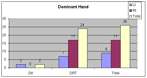
There is a relationship between dominant side and injury. 17 out of 26
had right side dominance (73%). Chi
Square 7.820534;
p value - 0.005166 (<0.05)
Dominant
Side on Side of Injury
Among
21 Patients under gone surgery within 48 hrs, average time for
surgery since injury is 24 hrs with a SD 7 hrs. For 4 patients
it is prolonged beyond 1 month due to late presentation.
Take the hypothesis that time interval between injury and surgery is in
the ratio 3:1:1 (<24 hrs, 24-48 hrs & >48 hrs)
Chi-square=0.410256 P-value=0.814543>0.05
| Duration |
15-20
minutes |
20-30minutes |
>30minutes |
| CN |
7 |
10 |
1 |
| Mini
open Radius |
0 |
3 |
0 |
| Mini
open R&U |
1 |
0 |
3 |
| Mini
open Ulna |
0 |
1 |
0 |
| Total |
8 |
14 |
4 |
| CN-
closed nailing |
|
Duration
of surgery & type of surgery |
Closed nailing was possible in 18 of 26 cases. Open reduction with
mini-incision was required in 1 ulna and 3 radial fractures
while in 4 cases both radius and ulna were opened. Among M/3,
Closed Nailing is commonly done (13/19). But there is no
statistically significant relation between site of fracture and
type of procedure like closed nailing or open reduction with
nailing.
Chi
square value - 0.28045; P value 0.869358 (>0.05)
The
average duration of surgery was 22.3 minutes. Only 4 cases
required more than 30 min for surgery. Out of 18 cases where
closed nailing was possible only one case took > 30 minutes.
In 8 cases where open reduction was required, only 3 cases had
operative time > 30 minutes.
Duration
of surgery is in the ratio 3:5:2 for <20 min, 20-30 min and
>30 min.
Using
chi square test chi-square=1.448639 with P value
.835699(>.05).
This
shows that operative time was not dependent on type of surgery
or open reduction did not cause extra operative time.
The
average hospital stay was 4 days with a range of 3 to 7 days.
Nearly 50% patients required only 3 days of Hospital stay.
Children with closed nailing were sent home on first
postoperative day.
Chi-square
0.929731; P value 0.999583
The
average union time was 6.7 weeks. Out of 18 cases where closed
Nailing was possible, 17 fractures united in 6 weeks. Only 1
case out of 8 where open reduction was required united by 10
weeks.
On
analysis with Grace and Eversmann outcome scoring out of 26
patients 20 had excellent outcome (77%), 5 had good outcome
(19.2%), 1 had acceptable outcome (3.8%) and there was no
patient with unacceptable outcome.
The
relation between fracture site and final outcome was not
statistically significant. Chi Square=0.000001 P value =0.999990
(>.05)
| |
Excellent |
Good |
Acceptable |
Unacceptable |
| L/3 |
5 |
1 |
0 |
0 |
| M/3 |
15 |
3 |
1 |
0 |
| P/3 |
0 |
1 |
0 |
0 |
| Total |
20 |
5 |
1 |
0 |
|
Functional outcome |
Closed
Nailing produce excellent result with 15 out of 18, Mini open
Radius + Ulna produce excellent results with 3 out of 4. But
Mini open Radius has 1 excellent outcome among 3 cases.
The outcome is excellent if the time for Surgery is <24 hrs in 15 out
of 20 (75%).There was no statistically significant relation
between final outcome and time interval between injury and
surgery.
Chi square value - 0.077235 P value –0.781081
We
had 7 complications in our study 2 olecranon bursitis, 2
superficial infection, 2 hypertrophied scars, and 1 ulna nail
back out. We had no non-union, malunion or limb length
discrepancies.
Conclusion :
The management of both bones forearm in children is critical for its
functional outcome at later age. There is an excellent
remodeling capacity of the pediatric long bones of forearm with
conservative treatment; however the rotational deformity still
persists. There is a big role of operative treatment in fracture
both bone of forearm in children.
The
fracture both bone forearms have been treated in past and till
date by closed reduction and above elbow pop cast
immobilization. This needs serial radiological reviews and
change of cast till the fracture consolidation. The good result
of union from conservative treatment have well proven[i]
but involves regular hospital visits, change of plaster cast,
care of cast, radiological reviews and at times operative
intervention if angulation is observed. Care of plaster cast in
pediatric age by the parents is very cumbersome job, which is
the mainstay of treatment for good functional outcome.
The
indication for operative intervention by plate osteosynthesis
for both bones forearm fracture is to give a better functional
outcome in displaced fractures. Despite of good results of rigid
plate fixation, long incision and extensive dissection is needed[ii]
[iii]
[iv].
This has a risk of infection, poor cosmetic, and need of implant
removal as second surgery, which again is an extensive
procedure. Furthermore stress shielding, plate breakage,
refracture are well known complication with surface fixation.
The
role of external fixation in fracture of both bone forearm is
limited only to open fractures and does not provide stable
fixation. There is also a concern of delayed union, pin tract
infection, joint stiffness and involves special care for
external fixator in children. On the other side, intramedullary
nail though biomechanically stable construct is not advised in
children with growing physis for the risk of damage to physis,
premature epiphysiodesis, or infection which are debatable.
In
1967 A K Talwalker[v]
designed square nail for forearm fractures of radius an ulna
using separate nails for treating adult forearm fractures. These
nails were used by various traumatologist in treating long bone
fractures with good results. Based on this concept surgeon from
France Nancy and Metz developed elastic titanium implant for
fixation of pediatric long bone fractures.
The
technique of intramedullary fixation of both bone forearm offers
several advantages of better anatomical-axial reduction, dynamic
stabilization, short hospital stay, less visits, with early
functional recovery, and simplified implant removal. The
technique of closed intramedullary nailing is minimally
invasive, respects biology of bone and soft tissues for better
and early bony union. Open surgery is not necessary except in
old or irreducible fracture. The minimal operative trauma,
undisturbed periosteal and endosteal vasculature, and axial
alignment maintained by nail which permits slight movement at
fracture site. All these factors favor rapid fracture union in
pediatric age group,
The square nail of Talwalker has a variable thickness
from 1.5 to 3 mm. preoperative evaluation was done and one nail
of adequate thickness was chosen to give a close fit to the
bone. Square cross section of nail helps to maintain rotational
alignment of radius and ulna by giving a close fit in medullary
canal. We had no displacement, no refracture as the implant
(square nail) was strong enough to hold the fracture till union.
The
closed reduction technique used for P/3, M/3, and L/3 fractures
was by application of traction to forearm in supination,
midprone and pronation of distal fragment. The reduction was
done under the control of image intensifier, by percutaneous K
wire joystick technique or by mini open surgery for radius (7
cases), ulna (5 cases). This mini-open surgery was required when
there was soft tissue interposition between fragments,
periosteal tube penetration and with uneducable fractures.
Although 8 cases required open reduction that has not affected
union time or functional outcome.
The
radial square nail was bent at the tip 10–20 degree for easy
entry at distal radial metaphysic for easy negotiation across
fracture site. This prebending was not a problem while
maintaining radial bowing.
The
point of insertion of radius is just proximal to the physis, a
small opening at distal metapysis (2 – 3 mm in length) and
enlarged by the curve of haemostat mosquito forceps. Ulnar nail
is inserted through the proximal ulna apophysis. As ulna square
nail is smooth at the tip it does not interfear with the growth
in children. The nails were passed short of 1 to 2 cm from
physis.
The average
time taken for surgery was 22.3 minutes, compared to 33. 5
minutes using titanium elastic nails by Richter et al 1998.
The
duration of hospital stay in our series was 3 days in 50 % of
our cases. All our patients were immobilized with long arm pop
slab for one week, followed by further immobilization in forearm
brace for 3 weeks. Those patients for whom mini open procedure
was done were immobilized for 6 weeks.
The
average time for fracture union in our series was 6.53 weeks and
average time for hardware removal was between 6 to 7 months.
None of our patients had non union. There was no limb length
discrepancy noted in our series. Infections were treated with
antibiotics. Olecranon bursa was excised at the time of implant
removal.
Cullen et al 1998[vii]
had a series of 20 children treated with Rush rods / Kirschner
wires with average time to fracture union was 10 weeks (range 6
– 22) without nonunion. Removal of hardware was at an average
16 weeks (range, 6-34). 17 patients had excellent results 2 had
good results and 1 had poor result. Complication occurred in 10
of 20 patients including 4 patients who required re-operation.
Eighteen complications occurred in 10 of the 20 children,
including hardware migration, infection, loss of reduction,
re-operation, nerve injury, significant decreased range of
motion, compartment syndrome, synostosis, muscle entrapment, and
delayed union.
Richter et al11
has reported 30 children (ages 4 to 14 years, 12 girls, 18 boys)
treated by titanium elastic nailing. 16 children were operated
immediately and 14 children late following failed conservative
treatment and fracture displacement. Average operative time was
33.5 minutes, and average duration of immobilization was 2 to 3
weeks. Implant removal was performed at an average 11.7 weeks
(range 8 – 26) after nailing. 24 children did not have any
discomfort, 3 had minimal discomfort, 3 had loss of supination
of 10 degree, and 2 had deficit in muscle strength.
The
delay in the hardware removal in our series was due to late
attendance as some of them waited for their school vacation for
the procedure.
We had complication of olecranon bursitis, and Ulnar nail backout as
some part of our ulnar nail was left out of bone for easy
implant removal. Olecranon bursa was removed at the time of
implant removal. No limb length discrepancies were noted as we
took measures not to use drill for making entry.
We had no nerve injury in our series as meticulous care was taken for
ulna proximal physis entry point such that the ulnar nerve is
kept medial by rolling the skin.
We
had no cross union/synostosis as we opened minimally by two
different incisions with a gap of 5 cm when mini open reduction
was done.
Reference :
-
Worlock
P, Stower M: Fracture patterns in Nottingham children.
JPediatr Orthop 6:656–660, 1986.
-
Landin
L A. Fracture patterns in children. Analysis of 8,682
fractureswith special reference to incidence, etiology and
secular changes ina Swedish urban population 1950–1979.
Acta Orthop Scand Suppl. 1983;202:1–109.
-
Mann
DC, Rajmaira S. Distribution of physeal and nonphyseal
fracturesin 2,650 long-bone fractures in children aged
0–16 years. J Pediatr Orthop. 1990;10:713–716.
-
Voto
SJ,Weiner DS, Leighley B. Redisplacement after closed
reduction offorearm fractures in children. J Pediatr Orthop.
1990;10:79–84.
-
Grace
T G, Eversmann W W Jr Forearm fracture; treatment by rigid
fixation with early motion. J Bone Joint Surg. Am,
1980;62:433-438
-
Creasman
C, Zaleske DJ, Ehrlich MG: Analyzing forearm fracturesin
children: The more subtle signs of impending problems. Clin
Orthop 188:40–53, 1984.
-
Wright
J, Rang M. Internal fixation for forearm fractures in
children. Techniques Orthop
1989;4:44-47.
-
VanderReis
WL, Otsuka NY, Moroz P, Mah J. Intramedullary nailing versus
plate fixation for unstable forearm fractures in children. J
Pediatr Orthop 1998;18:9-13.
-
Roy
DR, Crawford AH. Operative management of fractures of the
shaft of the radius and ulna. Orthop
Clin North Am 1990;21:245-250.
-
Talwalkar
AK & Talwalkar CA 1967. internal fixation of fractures
of radius and ulna in adults with Talwalkar intraamedullary
nails. Indian Journal of Orthopaedics Vol 1, No.1Jun 1967;26-30.
-
Richter,
Dirk M.D.; Ostermann, Peter A. W. M. D.; Ekkernkamp, Axel
M.D.; Muhr, Gert M.D.;Hahn, Micheal P. M.D. J Paed Ortop,
vol 18(4),july/aug 1998,457-461.
-
Cullen,Mark
C. M.D.;Roy, Dennis R. M.D,;Giza,Eric B. S.;Crawford, Alvin
H, M.D., F.A.C.S. J Paed Ortop, vol 18(1), jan/feb
1998,14-21
|

