|
Abstract:
We
present a case of fracture clavicle left treated with open
reduction and internal fixation with reconstruction plate and
screws in a 32 years old young man from a remote hilly village
of Eastern Nepal. He presented with chronic osteomyelitis of
clavicle with exposed implant in situ. This devastating
complication was noted after he had got treatment 14 months
back. This case is presented to share our bitter experience and
highlight one of the known but rare complications of this
fracture treatment.
J.Orthopaedics 2008;5(4)e11
Keywords:
Fracture clavicle;
chronic osteomyletis of clavicle;
treatment options of fracture clavicle
Introduction:
Clavicle fractures are common injuries,
representing about 4-10% of all adult fractures and 35-45% of
all fractures that occur in the shoulder girdle area. If these
fractures are classified into thirds, as proposed by Allman, the
most frequent site of injury is at the middle third (group I
fractures). These fractures account for approximately 72-80% of
all fractures of the clavicle. Approximately 25-30% of clavicle
fractures occur at the lateral clavicle (group II). Fractures of
the medial clavicle are quite rare, accounting for 2% of all
clavicle fractures in a recent epidemiological study by Nowak.
1
The clavicular
fractures can be treated by non operative or operative methods
according to its type, status of patient and the patients
choice. Nonoperative treatment of clavicle fractures consists of
sling support for 6 weeks. During this period, the patient does
perform pendulum exercises for shoulder motion and active range
of motion of the elbow and hand. After 6 weeks, the patient
begins passive assisted motion of the shoulder and progresses to
active range of motion as tolerated. Use of the sling may be
discontinued as pain allows.
Many techniques of
surgical fixation of clavicle fractures have been described in
the literature. When using plate and screw fixation to treat
clavicle fractures, the surgeon must remember that the hardware
will likely be prominent. Proper closure of these incisions is
imperative to decrease the risk of painful, prominent hardware
along with exposure late.
Case Report :
A 32 years old young man from remote hilly
village of Eastern Nepal presented to Department of Orthopaedics
B P Koirala Institute of Health Sciences Dharan Nepal with
complains of exposed left clavicle with implant in situ with
discharging sinuses on the operated site for open reduction and
internal fixation with reconstruction plate and screws from
fracture clavicle left 14 months back. He had no problems for 1
and half months after getting treatment of fracture clavicle
with operative intervention as mentioned above. He then
developed a discharging sinus over the site and was increased
gradually leading to spontaneous sloughing of skin exposing the
implant. Then screws were extruded leaving only two screws on
either side of plate. The proper Orthopaedic care was not
possible due to its unavailability along with his socio-economic
status and geo-political problems of the country. As soon as he
presented to our Institute, the removal of implant, debridement
of the wound and dressing of the wound was done in the priority
basis in the first stage. The wound was daily dressed till it
was covered by granulation tissue. Then split skin grafting was
done over the granulation tissue. With due course of time, the
wound was healed without further complications. At the end of 9
month from the time of debridement, patient had a good outcome
with full range of movement of the left shoulder.
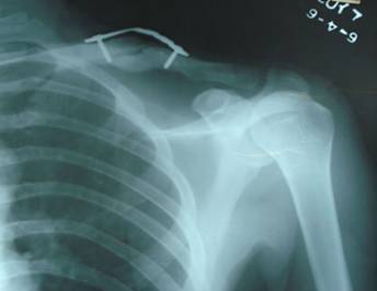
Fig A .Radiograph
showing implant failure for treatment of fracture clavicle with
plate
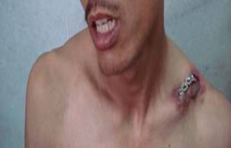
Fig B. patient with exposed implant on the clavicle with
osteomyelitis of clavicle
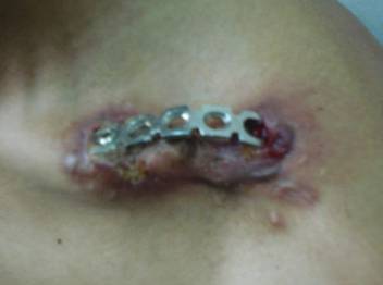
Fig C. Close up view of exposed implant and osteomyelitis
of clavicle
Discussion :
In neonates and children, these fractures
are very common and generally heal well. In adults, the force
required to fracture the clavicle is greater, healing occurs at
a slower rate, and risk of potential complications is higher.
The clavicle is the sole articulation of the shoulder girdle to
the trunk. It protects major underlying vessels, lung, and
brachial plexus. Displaced clavicle fractures can injure these
structures because of their proximity and sharp edges.
Extensive clinical
studies reported in the literature have indicated that
non-operative treatment is the treatment of choice for
clavicular fractures. It has also been suggested by some that
open reduction may contribute to the development of non-union.
From 1970 to 1978, twenty-five of approximately 800 patients
with a fracture of the clavicle were treated by open reduction
and internal fixation with a threaded intramedullary wire or pin
or with cerclage suture (one case). The patients' ages ranged
from thirteen to fifty-nine years. All fractures healed without
infection or migration of the pin. Based on this experience and
a review of the English-language literature, they concluded that
the indications for open reduction and internal fixation should
be: (1) neurovascular compromise due to posterior displacement
and impingement of the bone fragments on the brachial plexus,
subclavian vessels, and even the common carotid artery; (2)
fracture of the distal third of the clavicle with disruption of
the coracoclavicular ligament; (3) severe angulation or
comminution of a fracture in the middle third of the clavicle;
(4) the patient's inability to tolerate prolonged immobilization
(required by closed treatment) because of Parkinson's disease, a
seizure disorder, or other neuromuscular disease; and (5)
symptomatic non-union following treatment by closed methods. 2
Internal fixation of
the clavicle is rarely necessary. When it is warranted, the
clavicle's complex three-dimensional morphology and functional
anatomy, proximity to vital structures, and the multidirectional
biomechanical forces acting upon it place considerable demands
on any implant used for skeletal fixation. Mullaji AB et al
treated nine clavicles with the recently-introduced 3.5 mm low
contact-dynamic compression plate (LC-DCP). Surgery was
performed for symptomatic non-union in six patients, shoulder
dysfunction following a malunited fracture in one, for an open
fracture in one, and for an acute fracture associated with
brachial plexus injury in one. After an average follow-up period
of 17 months union was secured in each case. The advantages
afforded by the 3.5 mm LC-DCP in internal fixation of the
clavicle with its uniquely demanding anatomical and
biomechanical characteristics are discussed. 3
In a multicenter, prospective clinical
trial, conducted by Canadian Orthopaedic Trauma Society, 132
patients with a displaced midshaft fracture of the clavicle were
randomized (by sealed envelope) to either operative treatment
with plate fixation (sixty-seven patients) or nonoperative
treatment with a sling (sixty-five patients). Outcome analysis
included standard clinical follow-up and the Constant shoulder
score, the Disability of the Arm, Shoulder and Hand (DASH)
score, and plain radiographs. One hundred and eleven patients
(sixty-two managed operatively and forty-nine managed
nonoperatively) completed one year of follow-up. Most
complications in the operative group were hardware-related (five
patients had local irritation and/or prominence of the hardware,
three had a wound infection, and one had mechanical failure).
They concluded that operative fixation of a displaced fracture
of the clavicular shaft results in improved functional outcome
and a lower rate of malunion and nonunion compared with
nonoperative treatment at one year of follow-up. Hardware
removal remains the most common reason for repeat intervention
in the operative group. This study supports primary plate
fixation of completely displaced midshaft clavicular fractures
in active adult patients. 4
In another series of study done by
Poigenfürst J et al, there was no bony infection or
infected pseudarthrosis. Four clavicles refractured after
removal of the plate and five operations led to pseudarthroses
which were successfully treated by reoperation. Radiological and
clinical results in the majority of the re-examined patients
were excellent. Among total of 131 fractures of the clavicle
treated with plate and screws, such a devastating complication
as we found in our case, was not mentioned. 5
Ali Khan
MA etc al. reported treatment of twenty mid-clavicular
fractures by plate fixation. They mentioned that the technique
they used gave relief from pain within 12 hours and resulted in
bony union all cases. There were no such complications as they
reported. 6
23 fresh type II (Neer 1963) lateral
clavicular fractures were treated operatively. In 19 cases
fixation was done with two Kirschner wires, in four cases
plating was performed. The coracoclavicular ligament was left
unsutured. The average follow-up period was 4.5 (1-12) years. In
22 cases out of 23 the subjective outcome was good or
satisfactory. 22 fractures united and there were few
complications, but above mentioned complications were not found.
7
From the different large and small studies
from case series to multi-centric controlled trials, we
reviewed, did not mention such a devastating complication. Till
date there are controversies and debates on the treatment of
fracture clavicle, though we mostly observe its acceptable
alignment and union without specific interventions. We,
therefore, recommend conservative treatment of fracture clavicle
in our setup with geo-socio-economic conditions.
Reference :
- L Joseph Rubino. Clavicular fractures.
http://www.emedicine.com/orthoped/topic50.htm
- Zenni-EJ Jr; Krieg-JK; Rosen-MJ. Open reduction and
internal fixation of clavicular fractures J-Bone-Joint-Surg-Am.
1981 Jan; 63(1): 147-51
- Mullaji AB. Jupiter JB. Low-contact dynamic compression
plating of the clavicle. Injury. 1994 Jan 25(1):41-5.
- Michael D. McKee et al. (Canadian Orthopaedic Trauma
Society) Nonoperative treatment compared with plate fixation of
displaced midshaft clavicular fractures. A multicenter,
randomized clinical trial.
J Bone Joint Surg Am. 2007 Jan;
89 (1):1-10.
-
Poigenfürst J,
Rappold G,
Fischer W. Plating of fresh
clavicular fractures: results of 122 operations.
Injury. 1992;23(4):237-41
-
Ali Khan MA,
Lucas HK. Plating of fractures
of the middle third of the clavicle.
Injury. 1978 May;9(4):263-7.
-
Eskola A,
Vainionpää S,
Pätiälä H,
Rokkanen P.
Outcome of operative treatment in fresh lateral clavicular
fracture.Ann
Chir Gynaecol. 1987; 76 (3):167-9.
|

