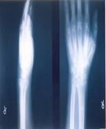
Out of 95 cases, 52 were male and 43 were female. Average age of
patients was 45.3 years ±7.5 years ranging from 18-83 years. 49
were having right sided and 46 were left sided fracture.
According to Frykmanns13 classification (1967), 45
cases were of grade I, 33 of grade II, 12 of grade III, 3 of
grade IV, 2 of grade VI and. Mode of injury in al the cases was
fall on the outstretched hand. All the cases treated in our
study presented to us within 1-8 days with mean of 1.80
days.
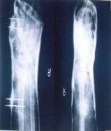
GROUP-A: 25 CASES
Functional result: excellent in12 (48%) and good in13 (52%)
Radiological results: excellent in 0, good in 13(52%) and poor
in 12(48%) cases
Mean differences in values of various parameters at 6th
week postop on affected side and normal side parameters
Radial
length-4.44mmwith SD-4.421 (normal-11.88, SD-1.36)
Radial angle-8.44 degree with SD-5.151 (normal-23.37, SD
-2.553)
Volar tilt-23.63 with SD- 14.872 degree (normal- [-7.0],
SD-7.004)
In 3 cases > 30 degrees of restriction of motion occurred, in 10
cases 15-30 degrees, in12 cases it was <15 degrees.5 cases were
having pain during heavy work. Grip strength was between
80-100%in all cases. Mild to moderate deformity was present in
23 cases with gross deformity in 2 cases.
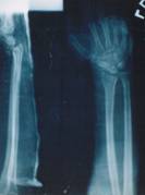
GROUP-B: 25 CASES
Functional results- excellent in 21(84%) cases, good in 4(16%),
poor in none
Radiological results- excellent in 20(80%) cases, good in
5(20%), poor in none Mean differences in values of
various parameters at 6th week postop on affected
side and normal side
parameters
Radial length-1.52mm withSD-0.714 (normal 11.88, SD-1.36)
Radial angle-2.98degree with SD -4.876(normal-23.37, SD-2.553)
Volar tilt- 9.88degree with SD -7.65(normal-[-7], SD-7.04)
In all cases, range of motion at wrist and forearm was nearly
full and painless with <10 degree of terminal restriction. Grip
strength was >95% in comparison to normal side in all cases. 1
case has shown slight widening of wrist. 1case had Sudecks
Osteodystrophy.
GROUP-C: 25 CASES
Functional results-excellent in 23(92%) cases, good in 2(8%),
poor in none
Radiological results- excellent-22 (88%) cases, good in 3(12%),
poor in none
Mean differences in values of various parameters at 6th
week postop on affected side and normal side parameters
Radial length-1.32mm withSD-2.260 (normal 12.08, SD-1.824)
Radial angle-2.68degreewith SD -5.037(normal-24.96, SD-5.713)
Volar tilt- 8.28degree with SD -7.895(normal-9.28, SD-8.028)
After a period of follow up the appearance of wrist was
identical to normal in 23 cases. 2 cases were having slight
widening. 23 cases were having range of motion with <10 degrees
restriction while 2 was having > 20 degree of restriction of
supination.Grip strength was >95% of normal.
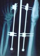
GROUP-D: 20 CASES
Functional results-excellent in 12(60.00%) cases, good in
6(30.00%), poor in 2 (10.00%)
Radiological results- excellent-60.0%cases, good in 30%, poor
in 10%.
Mean differences in values of various parameters at 6th
week postop on affected side and normal side parameters
Radial length-1.48mm withSD-2.260 (normal 12.08, SD-1.824)
Radial angle-3.86degreewith SD -5.037(normal-24.96, SD-5.713)
Volar tilt- 16.28degree with SD -7.895(normal-9.28, SD-8.028)
After a period of follow up the appearance of wrist was
identical to normal in14 cases. 4 cases were having slight
widening and 2 had obvious widening with ulnar styloid
prominence (these cases had gross displacement of reduction at 3rd
follow up week, we removed the distractor, did osteoclasis and
applied cast). 14 cases were having range of motion with <10
degrees restriction while 4 were having 10-20 degree of
restriction of supination and 2 was having restriction more than
20 degrees in supination and radial deviation. Grip strength was
>95% of normal in 18 cases while 2 had 85-95% of grip. 1 case
had Sudecks Osteodystrophy.
Discussion :
There are a lot of controversies whether anatomical reduction of
distal radial fractures is essential but there is no controversy
that maintaining satisfactory reduction is often difficult by
simple plaster cast. Bacorn and Kurtzke14 (1953)
analyzed the results of 2000 Colles fractures and they observed
that the poor functional results were directly related to the
degree of radiological deformity secondary to loss of position
at the fracture site in the plaster cast. Frykman13
(1967) also observed the same in his study.
There is no controversy that maintaining satisfactory reduction
in Colles fracture treated by simple method is often difficult.
Bacorn and Kurtzke14 (1953) and Frykman (1967) both
reported that lasting disability is greater in patients with
severe residual deformity. Other workers like Cassebaum (1950)
15, Lidstrom (1959) 16, Sarmiento17
et al (1975&1980), Stewart et al (1984) 5 found a
correlation between the anatomical and functional results at
three months but Stewart et al (1984) 5 reported that
this correlelation was lost by 6 months.
This finding of Stewart et al (1984) is confirmed in the
present prospective study, in which it was found that in spite
of less satisfactory radio graphical results (excellent 59.65%,
good 26.81%, poor 13.54%), the functional (clinical) result was
(excellent 76.66%, good25.83 %, poor 2.5%) with one having poor
functional results using the Schecks (1962) grading system or
the evaluation of end results.
These Schecks systems includes all the parameters and details
of subjective, objective and radiological finding and have
graded them with appropriate scoring system by which it becomes
very easy to obtain the functional and radiological results. The
same basis has been used for the comparison of the functional
and radiological results obtained in various other series.
This and all other assessments based on radiological and
objective measurements following distal fracture of the radius
have several limitations.
An accurate radiological measurement depends on comparable
view. A little change in the angulations of the x-ray beam or
positioning of the patients considerably alters bony
relationships. Measurements of the range of motion of the wrist
joint were considerably between examiners and in the same
patients at different times of the day, depending on previous
activity.
Indeed the range of motion of the wrist joint does not
necessarily denote function. A stiff painless wrist is for more
functional than a painful mobile one. Assessment of the function
is the best indication of the final result and is of major
concern to the patient.
In
the recent years, achieving and maintaining anatomical reduction
to improve ultimate function have been widely advocated,
particularly for intraarticular fractures. In spite of this,
initial poor anatomical alignment and secondary displacement
have been frequently accepted with distal radial fractures.
Anatomical reduction is not difficult to obtain, but as a result
of comminution of distal end of radius, the fracture is unstable
in reduced position. Garland and Warley18 (1951)
reviewing the final position of Colles fracture treated with
reduction and below elbow plaster immobilization, noted that in
60% cases union had occurred in a position, typical of a fresh
unreduced Colle's fracture. Mal alignment of the radio carpal
and distal radio-ulnar joints is inevitable. Cooney et al19
(1979) reported that post-traumatic arthritis was the second
most common complication of Colles fracture leading to pain,
weakness of grip and limitation of motion. They attributed this
to malaignment of the sigmoid notch of the distal end of the
radius with the ulnar head because of radial deviation and
dorsiflexion of the distal fragment or due to inadequate
restoration of length to ensure the normal radio-ulnar
relationship.
In present series, radiological results in comparison to each
others are as given in the table:
|
Radiological results
|
Closed reduction & cast (%) |
Functional cast brace
(%) |
Pin plaster technique
(%) |
Ligamentotaxis
(%) |
|
Excellent |
0 |
80.00 |
88.00 |
60 |
|
Good |
52.00 |
20.00 |
12.00 |
30 |
|
Poor |
48.00 |
- |
- |
10 |
Over all functional result in different series are:
|
Functional results i |
Closed reduction & cast (%) |
Functional cast brace
(%) |
Pin plaster technique
(%) |
Ligamentotaxis
(%) |
|
Excellent |
48.00 |
84.00 |
92.00 |
60 |
|
Good |
52.00 |
16.00 |
8.00 |
30 |
|
Poor |
- |
- |
- |
10 |
Pin plaster and ligamentotaxis as a method for achieving the
reduction and maintaining it, eliminating the possibility of
secondary displacement and provides better radiological and
functional results compared to conventional cast immobilization
method.
Though the number of patients in the present series is small for
satisfactory statistical analysis, but it appears that initially
in the first week of fracture treatment, there was significant
association of better function with improved anatomical
position. But as the time advances, this correlation gradually
disappears and at one year, function is almost normal.
The better functional and radiological results in the present
study were due to, avoidance of secondary displacement and early
finger exercises due to rigid immobilization especially pin
plaster & ligamentotaxis by distractor thereby eliminating
complications of conventional plaster technique which are the
major advantages of pin plaster & ligamentotaxis.
Conclusion:
Based on clinical and radiological findings of fifty-five cases
treated by various methods, the following conclusion can be
drawn:
1.
It is easy to obtain reduction but difficult to maintain
it by simple plaster cast
2.
Union in displaced position leads to poor functional and
cosmetic results.
3.
Techniques used in-group B, C, &D is easy, requires
minimum skill and can be done easily in minor OT under brachial
block.
4.
There is no secondary displacement in-group C&D like in
A&B.
5.
Closed reduction and plaster cast as well as functional
cast brace should be used in stable non-comminuted extra-articular
fractures.
6.
Pin plaster & ligamentotaxis by distractor should be used
in unstable, comminuted and intra-articular fractures.
Ligamentotaxis has the advantage that it can be used in cases
where skin is having abrasions or lacerations, where pin-plaster
is not possible.
7.
Overall results were excellent in 71.00% cases, good in
26.50% cases, and poor in 2.50% cases. The poor and
non-excellent results had been noted in those cases in which the
volar tilt of distal radial articular surface couldnt be
maintained either because of comminution, loss of reduction or
improper case selection.
Reference :
1. Golden GN: Treatments and programs of Collies fracture.
Lancet 1; 511-14, 1963
2. Hollingsworth R, Morris J: The importance of the ulnar side
of the wrist in fractures of distal radius. Injury 7: 263-66,
1976
3. Linschied RL: kinematic consideration of the wrist. Clin
Orthop 202: 27-39, 1986
4. McQueen M, Casper J: Does the anatomic results affect the
final outcome? JBJS 70B: 649, 1988.
5. Stewart HD, Innes AR, and Burke FD: The hand complications of
Colles fractures J Hand Surg 10B: 103-06, 1985.
6. Palmer AK: The DRUJ: anatomy, biomechanics, and triangular
fibro cartilage complex abnormalities, hand clinic 3:31-40, 1987
7. Short WH, Palmer AK, Werner FW, et al: -A biomechanical study
of distal radial fractures. J hand Surg 12a: 529-34, 1987
8. Fernandez DL: Avant-Bras segment distal. In Muller ME,
nazarian s, Koch P: classification of AO des fractures des os
longs. Berlin, Springer-Verlag, 1987: 106-15
9. Jupiter JP, Lipton HA: Operative treatment of intraarticular
fractures of distal radius: the upper extremity pilon fracture.
Clin Orthop, under publication at that time
10. Taliesink TM, Watson HK: Midcarpal instability caused by
malunited fractures of distal radius. J hand Surg 9a: 350-57,
1984
11. Colles A: On the fracture of carpal extremity of the
radius. Edinburgh Med surg J10: 182-86, 1814.
12. Smith RW: Treatise on fracture in vicinity of joints and
certain form of accidental and congenital dislocations. Dublin,
Hodges & Smity, 1854
13. Frykman GK: - Fractures of the distal radius including
sequelae ----shoulder hand finger syndrome. Disturbance in DRUJ
and impairment of nerve function: a clinical and experimental
study. Acta Orthop Scand Suppl. 108: 1-155, 1967
14. Bacon RW & Kurtzke JF: -Colles fracture, a study of two
thousand cases from the Newyork state workmans compensation
board. JBJS, 35-A: 3; 643-58.
15. Cassebaum WH: Colles fracture, a study of end results. JAMA,
143:963.
16. Lidstram A: Fractures of the distal end of the radius: a
clinical and statistical study of end results. Acta Orthop Scand
Suppl-41
17. Sarmiento A, Pratt GW, berry mc et al: -Colles fracture,
functional cast bracing in supination. JBJS, 57-A: 311
18. Gartland JJ, Werley et al: -Evaluation of healed Colles
fractures. JBJS, 33-A: 895
19. Cooney WO, Linscheid RL, Dobyn SJH: - External pin fixation
for unstable Colles fractures. JBJS. 6-A: 6; 840-45.