Isolated fractures of Trochlea are extremely rare. Very few
authors report having seen these fractures. Laugier is credited
with its original description in 1853.
The anatomy of the Trochlea probably contributes to its rarity
as compared to the capitellum. The Trochlea is situated deep
within the elbow joint and thus protected from direct injury.
The transmitted force of the ulna tends to produce a wedging
action than a tangential shearing force. The rarity of this
fracture is likely to cause misdiagnosis and improper
management.
We report a case of isolated fracture of the Trochlea sustained
by a patient following a road traffic accident. This case is
presented for its rarity and outcome.
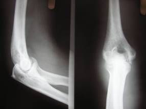
Isolated fracture of the Trochlea is a rare entity. Only
a handful of cases have been reported in literature; in our
review of literature we came across only five cases1,2,3,4,5.
Stimson has credited Laugier with its description in 1853, hence
it is also known as Laugiers fracture6.
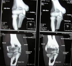
CT scan
showing the fracture
The fracture was exposed through a
medial approach to the elbow; the ulnar nerve was identified and
protected. The fracture was reduced and temporarily fixed using
Kirschner wires. The fracture reduction was checked using image
intensifier and was fixed using two 4 mm partially threaded
cancellous screws.
Post operatively the limb was immobilized in an above elbow
plaster slab for ten days and active flexion-extension exercises
begun in a functional brace. The brace was removed after four
weeks.
Patient was regularly followed up once in two weeks till the
fracture healed. Check radiographs were obtained at the tenth
post op day and at the end of six weeks. After six weeks
radiograph showed no loss of reduction with healing of the
fracture. At this stage, the elbow was found to be stable with
flexion 200 -1200 which was painless. The patient was followed
up till 1 year. At the last follow up, the patient was pain
free, having returned to his pre-injury occupation with full
elbow movements and no evidence of elbow instability. The
functional result was excellent according to functional rating
scale of Broberg and Morrey7.
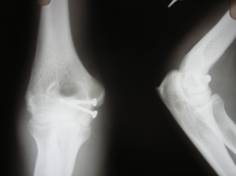
Post
operative X ray
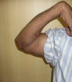
flexion at
the end of six weeks
Discussion :
Fracture of the Trochlea has been previously described as
part of the more complex fractures of distal end of humerus and
fracture dislocation of elbow 8; however it has been
very rarely reported as an isolated injury1,2,3,4,5.
The anatomy of the Trochlea probably contributes to the
rarity of this type of injury; it is deeply situated in the
elbow joint and thus protected from direct injury9,10.
The transmitted force of the ulna tends to produce a wedging
action than a tangential shear force10. Worrel
attributed the cause of an isolated trochlear fracture to a
force transmitted from the palm of the hand through the ulna to
the trochlea following a fall on the outstretched hand with the
elbow extended5.
As the fracture involves the osteochondral part of the distal
humerus it may be difficult to assess accurately on standard
radiographs11. It has been stated that, the one
finding that should lead the surgeon to suspect a fracture of
the trochlea is a fragment lying on the medial side of the joint
just distal to the medial epicondyle12. Two
modified lateral views have been described for better assessment
of complex elbow injuries. A radial head capitellar view might
be required to exclude injuries to the coronoid and type II
fractures of the capitellum.13
A coronoid trochlear view delineates the trochlea and coronoid
free of overlap of other bones.14
When in doubt a CT scan is helpful for delineating the extent
and type of fracture more accurately.
The operative treatment of these fractures may be equally
difficult given the limited amount of sub chondral bone
available for stable internal fixation of the fracture fragments8,
11.
Failure to reduce this fracture anatomically may
adversely affect not only the arc of flexion and extension of
the elbow but also the intrinsic stability of the elbow provided
by the Trochlea- olecranon articulation10, 12.
Conclusion:
Sonography is a useful
modality in diagnosis of severe CTS. We found out a decrease in
anteroposterior diameter of carpal tunnel in severe cases of
this disease. A finding which has not been considered
previously. This finding of narrow carpal tunnel may be
considered as a predisposing factor in severe CTS. To evaluate
the
US
results in mild to moderate disease, more study will be needed.
Reference :
1. D.A. Foulk, P.A. Robertson and L.A. Timmerman, Fracture of
trochlea, J Orthop Trauma 9 (1995) (6), pp. 530532.
2. Kaushal, R. , Bhanot, A. , Gupta, P.N. Isolated shear
fracture of humeral trochlea (2005) Injury Extra
3. Kwan, M.K. , Khoo, E.H. , Chua, Y.P. Isolated displaced
fracture of humeral trochlea: A report of two rare cases (2007)
Injury Extra
4. A.Oberstein, K.F. Kreitner, A. Lowe and I. Michiels, Isolated
fracture of trochlea humeri following direct elbow trauma,
Aktuelle Radiol 4 (1994) (5), pp. 271273
5. R.V. Worrel, Isolated displaced fracture of the trochlea, NY
State J Med 71 (1971) (19), pp. 23142315
6. Stimson L A, A treatise on fracture, Philadelphia, Henry C
Lea & co, 1890
7. M.A. Broberg and B.F. Morrey, Results of delayed excision of
radial head after fracture, J Bone Joint Surg 68(A) (1996), pp.
669674.
8. Gejrot,W,: On intra-articular fractures of the capitellum and
Trochlea of the humerus with special reference to the
treatment.Acta Chir Scandinavica,71:253-270,1932.
9. Bryan, RS ; Fractures about the elbow in adults, Instr Course
Lect, 30: 200 223, 1981
10. Eliason, EL & North, JP: Fracture about the elbow, Amr. J.
Surg, 44: 88 99, 1939.
11. Mckee,MD;Jupiter,JB;Bamberger,HB: Coronal shear fracture of
the distal end of the humerus.J. Bone and Joint
Surg.78,49-54,1996.
12. Smith, FM; Surgery of the elbow ; 2nd ed, Philadelphia; W B
Saunders 1972
13. A.Greenspan, A. Norman and H. Rosen, Radial head-capitellum
view in elbow trauma: clinical application and
radiographic-anatomic correlation, Am J Reontgenol 143 (1984)
(2), pp. 355359
14. J.C. Guilbeau, M.M. Monelhi and H. Nahum, Modified profiles
of the elbow in traumatology: the value of radial head-capitellum
view and a new coronoid-trochlea view, J Radiol 65 (1986) (5),
pp. 439444