Results :
In vitro study
Among the 15 cadaver tendons tested 5 each
were with Modified Kessler, Strickland and Four strand cruciate
techniques. The data was collected in the form of Force to
produce 2 mm gap formation at repair site; Maximum failure force
and Mode of failure.
(Table 1)
|
|
Force to produce 2 mm gap (Kg) |
Maximum failure force (Kg) |
|
Modified Kesslar |
0.8 |
0.9 |
|
Strickland |
0.9 |
1.0 |
|
Four strand cruciate |
1.8 |
2.12 |
In Modified Kessler technique, less than 1 kg of mean force
produced both 2 mm gap at repair site and failure of repair
(Maximum failure force).
In Strickland method, average 1 kg of force produced
failure.
While in four strand cruciate technique, average force to
produce 2 mm gap was 1.8 kg and 2.12 kg for repair to fail which
was almost double amount of force as compared to other 2
methods.
The
average force to produce 2 mm gap and maximum failure force in
Modified Kessler and Strickland is almost equal. The paired
studentst test was used to study the amount of increased
force from 2 mm gap formation to maximum failure force. We found
that there was no statistical significant increase in force in
these 2 groups.
Mod. Kessler t value = 1.952
p value = 0.2662 (p > 0.05)
Strickland - t value = 1.4142
p value = 0.1950 (p > 0.05)
But in case of four strand cruciate
method, this increase in force between 2 mm gap formation and
maximum failure force was 0.3 kg which was statistically
significant.
t value = 4.003
p value = 0.0039 (p < 0.05)
Among 15
cases, 60% (9/15) repairs failed by pullout of sutures from
tendon ends and 40 %( 6/15) failed by breakage of sutures. In
four strand cruciate group 80% (4/5) failed by suture breakage
while 20% failed by pullout. In other 2 methods, 80% repairs
failed by pullout
(Table
2)
|
|
Pull out |
Suture Breakage |
|
Mod. Kessler |
4 |
1 |
|
Strickland |
4 |
1 |
|
Four strand cruciate |
1 |
4 |
In Vivo study
There were 18 patients with lower limb tendon injuries. Total 19
tendoachilles tendons were repaired (1 patient had B/L TA
injury). The age distribution of patients was uniform as shown
by kolmogorov test
(Table 3)
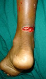
The ratio of injuries according to occupation was laborers
31.57%, carpenters and sedentary workers as 10.53% each. The
other occupations like students, housewives were 47.47%. 36.84%
injuries were due to closet injuries (foot slips and traps in
Indian closet, repeated attempts to remove foot causes sharp
laceration of tendoachilles). Injuries by knife accounted for
26.32% while RTA caused 15.79% injuries. The other modes of
injuries like assault, attrition were seen in 21.05% cases.
(Table 4)
|
Age |
No of Pts. |
|
<30 |
5 |
|
30 40 |
5 |
|
40 50 |
4 |
|
> 50 |
5 |
(Table 5)
Seven out of
19 tendoachilles injuries were due to closet injury (36.84%)
while it was injured by RTA in 26.31% and by knife in 15.78%.
The sharp cut injuries were seen in 13/19 tendons (68.42%),
while 3/19 were frayed (15.79%) and 3 were lacerated
(15.79%).All the injuries by knife were sharp while in closet
injury 5/7 were sharp (71.43%) and 2/7 were lacerated
(28.57%).The 14/19 tendons were right sided (73.68%) while 5/19
were left sided (26.32%).
(Table 6)
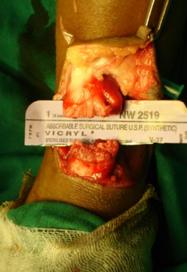
The average
operative time for tendoachilles repair was 32.81 minutes with
S.D. ± 7.52 min. Average operative time for sharply cut tendons
was 30 min. S.D. ± 5.77 min while for frayed tendons it was
43.33 min S.D.± 5.77 min. the lacerated tendons required 31.66
minutes with S.D. ± 2.89 minutes. The operative time difference
between these groups was statistically not significant.(t =
0.6407, p value = 0.48)
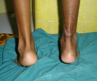
The
plantarflexion movement at ankle > 40° at 3 months was seen in
8/19 cases (42.10%) while planterflexion at 6 months was > 40°
in all patients.
At 3 months there were 10 patients with dorsiflexion at
ankle < 10° and 9 patients with dorsiflexion > 10°. At 6 months
all 19 limbs had dorsiflexion at ankle > 10°.
The average AOFAS score at 3 months was 76.25 with S.D. ±
7.96 and at 6 months was 87.125 with S.D. ± 3.81. The increase
in AOFAS score from
3 to 6 months was 11.125
(Table 7)
|
|
3 months |
6 months |
|
Plantar flexion >40° |
8/19 patients |
19 |
|
Plantar flexion <40° |
11/19 patients |
0 |
|
Dorsiflexion >10° |
9/19 patients |
19 |
|
Dorsiflexion <10° |
10/19 patients |
0 |
|
AOFAS score avg |
76.25 |
87.125 |
In patients with age less than 30 years, average AOFAS
score at 3 months was 79.2 which improved to 89.8 at 6 months.
In age group of 30 40 years AOFAS score improved from 77 to
87.4 while in 40 50 years group it improved from 77.25 to
86.5. In patients with age > 50 years score of 76.2 improved to
86.2. Patients with < 30 years age had better AOFAS score at 3
and 6 months than others but improvement in score from
3 to 6 months
was same in all age groups (10.8).
(Table 8)
Two patients had superficial infection. In one case it was
associated with skin dehiscence and slough. Both the cases
responded to oral antibiotic course and dressings. Patient with
skin dehiscence had AOFAS score 82 which was less than average.
Three patients had adhesions in which
2 were unable to use Indian closet and had below average AOFAS
score. Only 1 patient in our series had sural nerve paresthesia.
Discussion:
Since 1970 the management of tendon
injuries is revolutionized due to immediate tendon repair and
post repair motion protocols. Over these years many new suture
designs, methods has been developed to increase strength and gap
resistance of tendon repair techniques. They have permitted more
aggressive post repair motion protocols and hence the global
improvement in results. In spite of all these advances, there is
no consensus over the gold standard or an ideal tendon repair
technique.
A four strand cruciate design for
tendon repair as described by McLarney3
and Strickland seems to be the near ideal suture technique. A
review of literature shows lack of comprehensive clinical study
in tendoachilles repair with this technique. Hence this study
was performed.
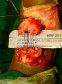
In our study, 15 cadaver tendons were
repaired by 3 methods viz. 1) Modified Kessler, 2) Strickland
and 3) Four Strand Cruciate and the tensile strength of repairs
was judged by linear force. For four strand cruciate the force
to produce 2 mm gap was average 1.9 kg which was double the
force required for other 2 methods. The maximum failure force
was also double as compared to other methods. The attributed
reasons for high tensile strength of four strand cruciate was 4
number of strand while other methods had only 2 strands. Lotz[i]
in an analytical model proved that four strand cruciate has more
tensile strength. In repairs with same number of strands, design
of core repair determines the strength.
The mode of failure depends on core suture design and
whether suture is locking or grasping type. The locking
configuration is one in which the transverse component is passed
superficial to the longitudinal so that suture passes around a
bundle of tendon fiber and usually prevents pullout. In grasping
type, the transverse component passes deep to longitudinal one
so that suture does not pass around or lock and is more prone
for pull out.
In our study, all repairs were grasping
type so the expected result was pull out of sutures at maximum
failure force. Though 80% (4/5) of modified Kessler and
Strickland sutures failed by pull out, surprisingly 80% (4/5
repairs) of Four Strand Cruciate failed by suture breakage.
Though a grasping type repair four strand cruciate exhibits
strong pull out resistance due to 4 strands and cruciate design.
The locking designs are known for more
gliding resistance and adhesions than grasping types. The
location of knot in four strand cruciate is away from the repair
site. This helps in decreasing bulk at repair site and assuring
perfect apposition of tendon ends. More over 4 grasping sutures
on surface may make this design more prone for adhesions. But in
our cadaver study we were not able to measure and compare
gliding resistance due to lack of costly devices like Load
Transducers.
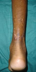
In lower limb, tendoachilles were the commonest tendon
injured. All the patients had minimum arc of movement from 10°
dorsiflexion to 40° platarflexion at ankle. Even the AOFAS score
showed good improvement between 3 to 6 months with 6 months
score being average 87.125 indicating good functional outcome.
These good results in tendoachilles were attributable to
immobilization of ankle in neutral position. Even the period of
immobilization was reduced to just 4 weeks contrary to 8 weeks.
Due to this strong repair we were able to mobilize patients
early with active motion protocols. Khan et al[ii]
in Cochrane review reported rerupture rate of 2.3% to 5% after
tendoachilles repairs while in our series there was no case of
rerupture. Even the skin dehiscence was seen only in single case
and 2 patients had superficial infection without any functional
problem. Overall in tendoachilles repair four strand cruciate
was found to be a good technique with sustainable persistent
good results without any major complication.
Conclusion:
The purpose of this study was to compare biomechanical
properties of Four Strand Cruciate technique with other
established techniques and to evaluate clinical results of this
technique.
-
Four
Strand Cruciate has significant high tensile strength and gap
resistance than other two strand techniques which can allow us
to use aggressive post repair rehabilitation protocols.
-
The Four Strand Cruciate with its peculiarities causes fewer
complications like reruptures and adhesions.
-
The ease of placement and less time for repair with above
properties make it near ideal tendon repair technique.
-
There is no significant correlation between tendon ends,
operative time and functional outcome of tendons repaired.
-
There are significantly good results in patients less than 30
years with respect to functional outcome irrespective of type of
tendons involved.
-
This technique gives confidence to the surgeon to start with
aggressive rehabilitation protocols to achieve good functional
results.
-
The incidence of complications like reruptures, adhesions, skin
dehiscence is very less.
Reference :
1. Maffuli N. Rupture of the Achilles tendon. J Bone Joint Surg
1999;81-A(7):1019-36
2. Cetti R, Christensen SE. Surgical treatment under local
anaesthesia of Achilles tendon rupture. Clin Orthop
1983;173:204-208.
3. Cetti R, Christensen SE, Ejsted R, et al. Operative versus
nonoperative treatment of Achilles tendon rupture. A prospective
randomized trial & review of literature. Am J Sport Med
1993;21:791-799.
4. Savage R. In vitro study of a new method of flexor tendon
repair. J Hand Surg [Br] 1985;10:135-141.
5. McLarney E, Hoffman H, Wolfe SW. Biomechanical analyses of
the cruciate four strand flexor tendon repair. J Hand Surg[AM]
1999;24:295-301.
6. Akoi M, Manaske PR, Pruitt DL, Kubota H, Larson BJ. Work of
flexion after flexor tendon repair with various suture methods.
A human cadaveric study. J Hand Surg[Br] 1995;20:310-313.
7. Lotz JC, Hariharan JS, Diao E. Analytical model to predict
the strength of tendon repairs.J.Orthop Res.1998;16(4):399-405
8. Khan RK, Fick D, Brammar T. Interventions for treating acute
Achilles tendon ruptures. Cochrane Database Syst Rev
2004;3:CD003674.