|
Mohammed
Benzagmout *, Saïd Boujraf **, ***, Taoufik Harzy#, Khalid
Chakour *, Mohammed El Faïz Chaoui *
*
Department of Neurosurgery,
University
Hospital
Hassan II,
Fez-
Morocco
.
**Department
of Biophysics and Clinical MRI Methods Department, Faculty of
Medicine and Pharmacy,
University
of
Fez
***Department of Radiology,
University
Hospital
Hassan II,
Fez-
Morocco
.
# Department
of Rheumatology,
University
Hospital
Hassan II,
Fez-
Morocco
Address for Correspondence:
Associate
Prof. Saïd Boujraf
Department of Biophysics and Clinical MRI Methods Department
Faculty of Medicine and Pharmacy,
University
of
Fez
BP. 1893; Km 2.200,
Sidi Hrazem Road
;
Fez
30000;
Morocco
Phone: 00 212 67 780 442, Fax: 00 212 35 619 321
E-mail: sboujraf@hotmail.com
|
|
Abstract:
Discal
calcification in childhood is rare. We report a 12-year-old boy
who presented an acute low-back pain, right L5 hyperalgic
sciatica with a history of increasing paresthesia. CT scan
demonstrated a postero-lateral calcified disc herniation at the
L4-L5 level. The patient was operated and successfully
recovered. Clinical presentation, neuroimaging findings and
treatment modalities of this phenomenon are discussed.
J.Orthopaedics 2007;4(3)e24
Keywords:
lumbar disc
herniation, intervertebral disc calcification, infant, surgery
Introduction:
The surgery of lumbar disc herniation is a relatively uncommon in
children. In published series, children generally constitute 0.5
to 3% of all patients surgically treated for lumbar disc
herniation [3, 4]. Moreover, calcifications of the
intervertebral disc occur rarely at childhood stage [13], and
most commonly involve the lower cervical spine [6]. It may be an
incidental finding, or associated to distinct clinical syndrome,
and it is rarely associated to neurological deficit [15].
In this paper, we present the case of a calcified lumbar disc herniation
revealed in a child of 12 years by hyperalgesic sciatica. The
patient was operated in emergency with a very good outcome.
Case
Report :
Our
patient was a 12 years old sportive boy, admitted at the
neurosurgical emergency department for a severe pain of the
lumbar spine. He reported low back pain, difficulty of walk,
typical right L5 sciatica and a history of increasing paresthesia
in his lower limbs. There was no history of trauma. These
symptoms occurred one month earlier and the patient has received
treatment consisting in analgesics, non- steroidal
anti-inflammatory drugs and muscle relaxant for a period of
three weeks without any improvement of his complaints. Moreover,
the patient reported an increasing severity of symptoms
justifying radiological exploration. At admission, the physical
examination revealed radiative spinal syndrome (muscular
tightness associated to antalgic attitude by trunk flexion,
Lasegue sign of 30° toward the right). However, the
neurological examination did not reveal any motor or sensory
deficits.
The
basic x-rays of the lumbar spine did not show any particular
signs. Whereas CT scan of the lumbosacral
junction
demonstrated a postero-lateral calcified disc herniation
localized at the L4-L5 level (Figure 1).
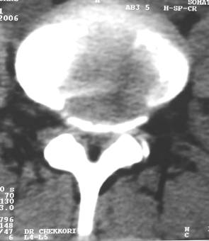
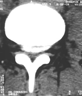
Figure 1: CT scan of the
lumbar spine in axial views at the level of L4-L5 intervertebral
disc (a) and the superior vertebral plateau of L5 demonstrating
a postero-lateral calcified disc herniation.
The
others lumbar intervertebral spaces were normal. We recommended
that the patient undergo L4-L5 discectomy. In surgery, we
discovered a calcified lumbar disc herniation at the level of
L4-L5 without clear conflict with the L5 nerve root. A bilateral
foraminotomy was performed and the patient showed spectacular
improvement of his symptoms after surgery. After two months, the
patient returned progressively to practice his favorite
preoperative sport and lifestyle.
Discussion :
Calcified intervertebral discs is very rare in children, it was firstly
described by Baron in 1924 [2], about 250 cases were previously
reported the literature [8]. In fact, the real incidence of this
pathology in children and adolescents is still controversial
according to the upper age limit of this population.
The etiology of disc calcification in children remains unclear [7, 15];
however, it is possibly different from the degenerative
calcification seen in adults. Trauma has often been implicated
as a predisposing factor since the calcification of disc in some
children was preceded by trauma [6, 12, 15, 17]. However,
preexisting disc calcification was also identified in some
children [14], and most patients have no history of injury.
Furthermore, disc calcification in newborns has been reported
[10]. Thus, it is difficult to determine the relationship
between trauma and disc calcification in children.
Indeed, several factors have been investigated, such as familial
predisposition, the presence of morphologic and functional
alterations, congenital malformations, growth disturbances, and
vertebral slipping epiphysis. Nevertheless, traumatism is often
mentioned as the primary causative factor [5].
The mean age at onset is about 7 to 8 years with a slight male
preponderance. The clinical presentation is variable. Most
patients have local symptoms including pain, muscular tension
and functional limitation [8]; some patients may have a fever
[9]. Rechtman et al. [14] classified the clinical symptoms of
disc calcification as disappearing, dormant and
silent types. The severity of the symptoms is often not
correlated with the radiographic findings [12], and the
calcification may be only an incidental finding [14, 16]. In
most cases, it has been accepted that disc calcification in
children is self-limiting and has an excellent prognosis [8].
However, herniation of calcified discs occasionally leads to
acute nerve-root or spinal cord compression urging a surgical
decompression [9]. This was the case of our patient who
experienced sudden hyperalgesic
sciatica without any history of symptoms.
Radiographically, the calcified discs are usually seen as a dense round
or oval mass within the nucleus pulposus. Both CT and MRI can
demonstrate an associated disc herniation [12]. The
calcification has been described as having low signal intensity
on magnetic resonance images [12], although hyper-intense discs
on the T1-weighted image are associated with calcification [1].
Disc calcifications are mostly found in the cervical spine with
a clear predilection for the C6-C7 level [8]. The thoracic area
is rarely involved [10] whereas the lumbar spine is
exceptionally affected [16]. Multiple disc involvement has also
been reported [8].
Conservative treatment is usually effective [17]. It allows the
disappearance of clinical symptoms in 70% of cases in parallel
to calcified disc vanishing. In fact, most of authors believe
that surgical intervention for the treatment of nerve-root or
spinal cord compression by a calcified disc is rarely indicated.
However, surgical intervention has been reported in some
patients with intractable pain and/or a progressive neurological
deficit [9, 15]. In our case, the intervention was justified by
the severity of the radiculopathy that was rebel for medical
treatment.
The prognosis of intervertebral disc calcification in children is
excellent [8]; and complications rarely occur [11].
Conclusion:
Disc calcification in childhood represents a rare entity. It is mostly
revealed by a characteristic acute spinal pain and usually
follows a benign course. CT scan is the exam of choice. It
allows to confirm the diagnosis and to exclude other diseases,
mainly bacterial spondylo-discitis. Surgery is rarely required and the prognosis is usually
excellent.
Reference :
-
Bangert AB, Modic MT, Ross JS, Obuchowski NA, Perl J, Ruggieri PM,
Masaryk TJ. Hyperintense disks on T1-weighted MR images:
correlation with calcification. Radiology.
1995; 195: 437-43.
-
Baron, A.: Uber eine neue Erkrankung der
Wirbelsaüle. Jahrb. Kinderh.
1924; 104: 357-360.
-
Beks JW, Weeme CAT. Herniated lumbar discs in teenagers. Acta
Neurochirurgica (Wien) 1975; 31: 195-199.
-
DeOrio JK, Bianco A. Lumbar disc excision in children and adolescents.
J Bone J Surg 1982; 64A: 991-5.
-
Epstein JA,
Epstein
NE
, Marc J, et al. Lumbar
intervertebral disc herniation in teenage children: recognition
and management of associated anomalies. Spine 1984; 9: 427-32.
-
Eyring EJ,
Peterson
CA
, Bjornson DR. Intervertebral-disc calcification in childhood: a
distinct clinical syndrome. J Bone Joint Surg Am. 1964; 46:
1432-41.
-
Gerlach R, Zimmermann M, Kellermann S, Lietz R, Raabe A, Seifert V.
Intervertebral disc calcification in childhood: A case report
and review of the literature. Acta
Neurochir (Wien) 2001; 143: 89-93.
-
Harvet G., De Pontual L., B. Neven, et al.Calcifications
discales de lenfant : à propos de deux observations et revue de
la littérature.
Archives de pédiatrie 2004 ; 11 : 1457-1461.
-
Li-Yang D, Hua Y, Qi-Rong Q. The Natural History of Cervical Disc
Calcification in Children. J. Bone Joint Surg. Am. 2004; 86:
1467-1472.
-
MacCartee CC Jr,
Griffin
PP, Byrd EB. Ruptured calcified thoracic disc in a child. Report
of a case. J Bone Joint Surg Am. 1972; 54: 1272-4.
-
Mahlfeld K, Kayser R, Grasshoff H. Permanent thoracic myelopathy
resulting from herniation of a calcified intervertebral disc in
a child. J Pediatr Orthop B 2002; 11: 6-9.
-
McGregor JC, Butler P. Disc calcification in childhood: computed
tomographic and magnetic resonance imaging appearances. Br J
Radiol. 1986; 59: 180-2.
-
Prescher A. Anatomy and pathology of the aging spine. Eur J Radiol.
1998; 27: 181-95.
-
Rechtman AM, Hermel MB, Albert SM, Boreadis AG. Calcification of the
intervertebral disk: disappearing, dormant and silent. Clin
Orthop. 1956; 7: 218-31.
-
Smith RA, Vohman MD, Dimon JH 3rd, Averett JE Jr, Milsap JH Jr.
Calcified cervical intervertebral
discs in children: report of three cases. J
Neurosurg. 1977; 46: 233-8.
-
Ventura N, Huguet R, Salvador A, Terricabras L, Cabrera AM. Intervertebral disc calcification in childhood. Int Orthop. 1995; 19:
291-4.
-
Wong CC,
Pereira
B, Pho RW. Cervical disc calcification in children. A longterm
review. Spine. 1992; 17: 139-44.
|

