|
Chowdhry
M*, Bismil Q, Blackburn SC, Little N, Ricketts DM
*Orthopaedic
department of Brighton and Sussex University Hospitals NHS
Trust
Address for Correspondence:
Majid
Chowdhry
12 Woodfield Grove, Streatham, London, SW16 1LR
E-mail: m.chowdhry@doctors.org.uk
Tel: 07920112030
|
|
Abstract:
Snapping
scapula is an uncommon symptom associated with shoulder pain and
reduced movement. Elastofibroma is a rarer cause of snapping
scapula.1 We describe a rare cause of snapping scapula: benign
elastofibroma at the inferior pole of the scapula. This was not
demonstrated by CT scanning but the lesion was successfully
treated by excision, and histology determined the diagnosis of
elastofibroma.
Keywords: snapping scapula, elastofibroma.
J.Orthopaedics 2007;4(2)e11
Introduction:
Snapping
or clicking beneath the scapula is an uncommon symptom. We
present an unusal cause: elastofibroma of the chest wall.
Elastofibromas are uncommon, benign, slow-growing connective
tissue tumours; and are often periscapular.
They rarely cause scapular snapping.1
Diagnostic difficulties arise in their illusiveness with the use
of CT scans. MRI has proven to be a much better form of imaging.
5
Case
Report:
 A
61-year-old right handed businesswoman presented with a 5 month
history of painful snapping of the left scapula. There was no history of trauma.
On examination scapulothoracic crepitus was observed.
There was a 4 x 4cm hard, smooth-surfaced lump at the lower pole
of the scapula, partially tethered to the underlying ribs.
The patient was generally fit and well, the general
examination was unremarkable and the right scapula was normal. A
61-year-old right handed businesswoman presented with a 5 month
history of painful snapping of the left scapula. There was no history of trauma.
On examination scapulothoracic crepitus was observed.
There was a 4 x 4cm hard, smooth-surfaced lump at the lower pole
of the scapula, partially tethered to the underlying ribs.
The patient was generally fit and well, the general
examination was unremarkable and the right scapula was normal.
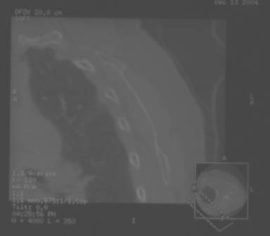
Fig.1
CT formatted saggital view of Left scapula
 Radiographs
of the scapula, including a scapular lateral view were normal.
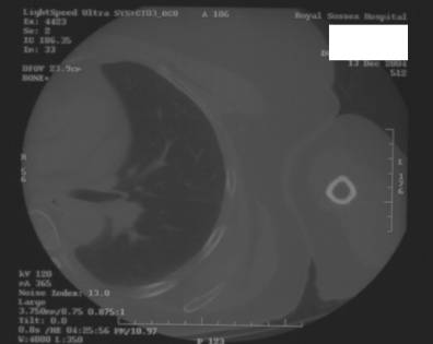
CT scan of the left shoulder was also normal (figure 1, 2). Radiographs
of the scapula, including a scapular lateral view were normal.
CT scan of the left shoulder was also normal (figure 1, 2).
Fig.2 CT
axial view of the inferior angle of the left scapula.
At
operation under general anaesthesia, an oblique incision over
the lower pole of the scapula was used to visualise the tumour.
The lesion was attached to the periosteum of the ribs and
the external fascia of the rib cage, and extended into
subscapular space. The tumour was excised and sent for
histology. The lump was composed of a heterogeneous mixture of collagen,
elastic fibres and fibroblasts; consistent with a diagnosis of
benign elastofibroma.
At
six week follow-up, the surgical site has healed and the patient
reported that the painful clicking had resolved. She had returned to work.
She has had no recurrent scapular clicking or pain.
Discussion :
The
causes of snapping scapula are classified as: deformity or
exostosis of the thoracic cage; an abnormal relationship between
the scapula and the thoracic cage secondary to poor posture;
lesions of the scapulothoracic muscles; lesions of the
subscapular bursae; and lesions of the scapula itself.
Lesions of the scapula include: increased anterior
inclination of the superior angle; osseous or fibrocartilaginous
nodules on the anterior aspect of the superior angle; tumours of
the anterior surface of the body of the scapula; Sprengel
deformity and malunited fractures.1
Elastofibroma
is an uncommon benign primary tumour of soft tissues2 .It
usually arises deep to the lower scapular pole;3 is often
bilateral;3 and consists of a mixture of collagen, elastic
fibres and fibroblasts.3
A
literature search revealed that elastoma of the chest wall has
been infrequently described as a cause of snapping scapula. Less
than 20 cases have been described in the literature2-4.
Literature
describes CT findings of elastofibroma as typically poorly
defined changes in the soft tissue, which although
characteristic of elastofibroma, dont always definitively
diagnose the lesion. MRI is more superior in this respect,
showing more clearly the same alternating high and intermediate
signal intensities on T1-weighted imaging that can be enhanced
by using gadolinium.5
Our case
presented with the triad of: lump at inferior scapular pole,
pain and snapping. The presence of the triad of inferior
pole lump, pain and snapping favour this diagnosis. CT may
not always demonstrate the lesion, in which case MRI is a better
investigation to perform
Reference :
-
Canale ST. Campbells
Operative Orthopaedics.
-
Majo J, Gracia I, Doncel A, Valera M, Nunez A, Guix M.
Elastofibroma dorsi as a cause of shoulder pain or
snapping scapula. Clin
Orthop Relat Res. 2001 Jul;(388):200-4.
-
Cohen I, Kolender Y, Isakov J, Chechick A, Meller Y. [Elastofibroma,
a rare cause of snapping scapula syndrome].
Harefuah.
1999 Oct;137(7-8):287-90, 350.
-
Nielsen T, Sneppen O, Myhre-Jensen O, Daugaard S, Norbaek J. Subscapular
elastofibroma: a reactive pseudotumor.
J
Shoulder Elbow Surg. 1996 May-Jun;5(3):209-13.
-
Abe S, Miyata N, Yamamoto Y,
Yamaguchi T, Tamakawa M. Elastofibroma Dorsi: CT, MRI and
pathologic findings. Plastic and Reconstructive Surgery. 1999
Dec;104(7):2121-2126
|

