Address for Correspondence:
Dr.Shailendra Khare
Orthopedic Surgeon, Central Institute of Orthopedics, Safdarjung Hospital & Associated V.M. Medical College, New Delhi 110015, India,
Phone : (C)0091-9810177245
Email: skhare245@rediffmail.com
Abstract:
Back ground: The present prospective study was carried out to evaluate clinical and radiological correction of the deformities in neglected, resistant and relapsed or recurrent clubfoot using Joshi’s external stabilizing system (JESS) and to compare the result of JESS Method with Standard one stage Posteromedial Soft Tissue Release (PMSTR).
Methods: In this prospective study 50 children were randomly divided into 2 groups. Group I comprised of 32 feet in 25 children who were subjected to Posteromedial Soft Tissue Release (PMSTR) using Turco’s Approach and Group II Comprised of 30 feet in 25 children who were subjected to differential distraction method by using JESS fixator. The results were evaluated using clinical and radiological criteria, as described by Simons in 1985, with some minor modifications.
Results – True radiological and clinical correction was achieved in group I in 68.75% cases. In group I the results were unsatisfactory clinically and radiologically when age of the patient at the time of surgery (PMSTR) was more than 12 months. Satisfactory radiological and clinical results were obtained in 73.33% in group II, where the mean age for surgical procedure was 2years and 4 months.
Conclusion- The children who came for treatment at a very early age and received regular treatment had overall better results. Children who were operated using Turco’s Approach (PMSTR) before 12 months had more satisfactory results but the results were unsatisfactory if this procedure was done in children over one year of age. The joshi’s external stabilization system is the method of choice in relapsed and neglected CTEV cases and the best results are obtained if done before 2 years of age.
J.Orthopaedics 2011;8(4)e8
Keywords:
CTEV, PMSTR, JESS
Introduction:
Talipes Equino Varus (“Club Foot”), a congenital deformity of foot, has continued to plague the medical profession since before the days of Hippocrates1. The deformity consists of hindfoot equinus, hindfoot varus, and forefoot varus. In developing countries, these children often report late for treatment either because of lack of medical facilities or due to ignorance. The management of such neglected or relapsed clubfoot unlike that of virgin cases is challenging because with time the deformities becomes fixed and the foot develops secondary adaptive bony changes. These feet usually are not amenable to correction by soft tissue release procedures alone and often need some bony procedures as well. However, bony procedures (closing wedge osteotomy, arthrodesis) lead to further shortening of an already smaller foot of CTEV.
For those cases presenting late or after relapse, there is a treatment option based on principle of Differential Fractional Distraction Histogenesis of G.A.Ilizarov2. However in order to successfully use the tensioned wires required for Ilizarov fixator, child must have attained the age of 3 years because prior to this age there is insufficient strength in cartilaginous tarsal bones. Dr. B.B. Joshi et al. based on the principle of Differential Fractional Distraction Histogenesis, has developed a simpler construct for the correction of clubfoot deformities known as JESS (Joshi’s external stabilizing system), which can be used even in children below three years of age because it doesn’t use tensioned wires3 . With JESS, the author has shown successful management of clubfoot deformities from the age of 3 months to late childhood.
The present study was undertaken with a view to assess the results of PMSTR and JESS fixator in neglected, resistant and relapsed or recurrent club foot and compare these procedures.
Material and methods
This prospective study comprising of 50 children, was conducted in the department of orthopedic surgery at DDU Hospital, New Delhi, between June 2004 and Dec. 2006. Only idiopathic clubfoots of neglected, resistant relapsed or recurrent type, in children less than four years, were included in this study.
The children were randomly divided into 2 groups
Group 1: It comprised of 32 feet in 25 children who were subjected to PMSTR using Turco’s Approach. These children were those who either failed to respond adequately to conservative treatment or had moderate to severe deformity when they reported 1st time for treatment, at about 6 – 9 months of age.
Group 2: It comprised of 30 feet in 25 children who were subjected to differential distraction method by using JESS fixator. These children had either resistant or rigid type of CTEV at about 1 – 4 year of age. Children with neglected or recurrent deformities, relapsed or recurrent deformities after previous unsuccessful soft tissue release were also included in this group.
All the selected patients in both the groups were subjected to a thorough general examination and a detailed local examination of foot. Routine investigations were obtained as a part of pre anaesthetic check up. Pre operative radiological assessment was done. Criteria for clinical and radiological assessment were same as described by Simon (1985) with slight modifications. Based on the pre-operative assessment, the surgical strategies were planned. I/V Antibiotics protocol included one dose prior to the surgery and continued for forty eight hours post operatively.
Group I protocol
All children’s were operated under general anaesthesia by the surgical procedure essentially same as described by TURCO’s, except that internal fixation with Kirschner wire was not done routinely.
Post-operatively, a cast was used extending from the thigh to the toes with the foot in neutral position and the knee flexed 60 degrees. The capillary circulation of toes was checked. The limb was kept elevated on a pillow postoperatively to avoid any swelling.
Patients were generally discharged within a week after the surgery.
First cast was charged after 2 weeks of surgery.
The second cast was changed after two weeks. At this time the Kirschner wires (if used) as well as stitches were removed.
Subsequently the patient was called at a weekly interval & cast was changed.
The last cast was removed at 8 weeks (number of cast applied post operatively were 5).
If however deep infection or major wound dehiscence were present then Kirschner wire were retained for a longer time and removed after 6 weeks. Casts were also applied for a longer duration. Usually 7 casts are applied and the last cast was removed at the 10th week.
After the cast removal patient were put on night splint and corrective shoes.
All patients were put on physiotherapy dorsiflexion / plantar flexion exercise and inversion / eversion exercises.
After two weeks of physiotherapy, when the foot had become pliable and mobile, a combined clinical & radiological assessment was done.
Group II protocol
All the children were operated under general anaesthesia by the surgical technique as described by Joshi et al. 3 [figure 1].
Distraction Protocol: On 3rd post operative day dressing of pin tract was performed. On 7th post operative day distraction was commenced. Medial tibio-calcaneal and the medial calcaneometatarsal distractor knobs were rotated 360 degree i.e. 90 degree rotation every 6 hourly and the lateral counterparts will be rotated 180 degree i.e. 90 degree every 12 hourly in the clockwise direction. Clinical & radiological correction was achieved approximately between 4 to 6 weeks. Following the correction the assembly was held in a static position for twice the period of distraction. The whole assembly was removed under general anaesthesia and plaster cast was given at the completion of static phase.
First three casts were molded above knee in maximum correction.
Change of cast with manipulation was necessary every two weeks for three months. The last cast was a below knee POP Cast for allowing the child to ambulate.
After 3 months of immobilization, Denis Browne Bar with attached open toe corrective shoe during sleeping hours for a maximum of one year was given. The child was also referred to physiotherapist for gait training and to take remedial steps to strengthen weak/ weakened muscles.
A complete clinical and radiological evaluation was done post operatively and during every follow up visit. X-rays were taken after surgery, during distraction phase and static phase, during follow up after three months and finally after one year to assess the correction of foot. All the patients were regularly assessed at 12 weeks interval.
Follow-up: In group I, three cases were lost after 12 weeks of follow up. Minimum follow up of cases in this group was 12 weeks and maximum being 90 weeks. The mean follow up was 42 weeks. In group II, minimum follow up was 24 weeks and maximum was 104 Weeks with mean follow up of 48 weeks.
Results:
There were 20 (80%) males and 5 (20%) females in group I. Of the total 32 feet in this group, twenty four feet (75%) were operated between the ages of 6 month to one year.
In Group II out of the 25 children, 14 (56%) were male and the remaining 11(44%) were female. Of the total 30 feet in group II, twenty nine feet (96.67%) were operated between the age of 1 year to 4 year of age . One case of failed previous surgery was operated at 11 months of age.
It was observed that unilateral deformity was more common than bilateral deformity in both the groups. Left side has slightly greater predominance than the right side in group I whereas right side was more commonly involved in group II. Associated congenital anomalies were not seen commonly. One case had Cleft Lip and Cleft Palate with excess Hairy Growth over the body. This patient had bilateral club feet; right side was operated using JESS FIXATOR and left side by TURCO’s procedure [figure2-5]. The average duration of plaster cast treatment was 29.75 weeks for group I, and 20.67 weeks for group II.
Table1: Showing Previous Treatment Records
Treatment prior to surgery |
Group I {PMSTR}
No. of Children (%) |
Group II {JESS}
No. of Children (%) |
No Treatment(Neglected cases) |
0 |
8(32) |
Irregular Conservative Treatment(resistant cases) |
8(32) |
7(28) |
Regular Conservative Treatment(Resistant cases) |
17(68) |
5(20) |
Previous Surgery(Relapsed cases) |
0 |
5(20) |
Eight children (32%) in group II had no cast treatment prior to surgery. In group II, five children who were operated previously had recurrence of deformity. They were operated with JESS FIXATOR.
Table II: Clinical Criteria for Assessment of satisfactory results*
S.N. |
CLINICAL CRITERIA |
SATISFACTORY* |
Group I ( n=32) |
Group II (n=30) |
1 |
Symptoms |
none |
None |
28 |
2 |
Appearance of Hindfoot |
0,1 |
25 |
25 |
3 |
Forefoot Adduction |
0,1,2 |
28 |
26 |
4 |
Foot knee malalignment |
0,1 |
25 |
|
5 |
** Functional weakness of calf |
None |
|
|
6 |
Movement at ankle joint |
Dorsiflexion > 100
Plantarflexion > 150 |
22(68.75%)
30(93.75%) |
26(86.67%)
30(100%) |
7 |
Movement at subtalar joint |
Present |
28 |
27 |
8 |
***Additional treatment |
Minor |
4 |
15 |
9 |
Complications |
Minor |
26 (81.25%). |
23(76.33%) |
*Anything other than this was considered as unsatisfactory and an unsatisfactory rating in any category was considered sufficient to grade the total clinical results as unsatisfactory.
** Functional weakness could not be assessed pre-operatively. However post-operatively,in Group I, it was found in 1 case out of 4 assessed. In group II, 4 cases out of 15 assessed had functional weakness.
***Child was referred to physiotherapist for gait training and to take remedial steps to strengthen weak/ weakened muscles. Most of the patients were explained about physiotherapy by the treating doctors.
Preoperative none of the feet in either group had a clinical satisfactory rating but after surgery a significant improvement was seen. Of the 32 feet subjected to PMSTR procedure it was observed that 22 feet (68.75%) were in the category of satisfactory rating where all the 9 clinical criteria were in satisfactory range and10 feet (31.25%) had unsatisfactory result. Of the 30 feet subjected to Differential Distraction Method using JESS FIXATOR it was observed that 23 feet (76.67%) were in the category of satisfactory rating, where all the 9 criteria were in satisfactory range, unsatisfactory result were seen in 7 feet (23.33%).
Complications:
In group I superficial wound infection and minimal wound sloughing were regarded as minor complications, and were seen in a total of 8 feet (25%) in group I. However deep wound infection and major wound dehiscence were seen in 6 feet (18.75%). Results of these six feet were rated as unsatisfactory.
In group II superficial wound infection / pin tract infection, swelling of the foot and pain with fixator were regarded as minor complications, and were seen in a total of 4 feet (13.33%). However superficial gangrene of toes due to foot plate pressure was seen in 4 cases and excessive valgus were seen in 3 feet (23.33%). No case of Osteomylitis or pin breakage was seen. These 7 cases were seen as major complications and results were rated as unsatisfactory.
Fig. 1(a)---------------------------- Fig. 1(b)
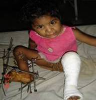 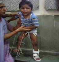
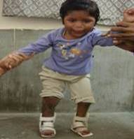 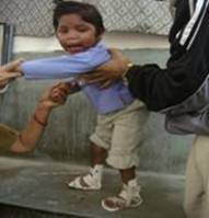
Fig. 1(c) ---------------------------- Fig. 1(d)
{3 years 4 months old female patient with cleft lip and cleft palate was operated for bilateral club foot. Right side by JESS Fixator & Left side using TURCO’s procedure. Fig.2(a): Post operative; Fig 2(b)Le:ft side is on splint and right side is maintained in static phase of JESS Fixator; Fig2(c) & 2(d) : Bilateral splint is applied}.
Table II: Radiological Criteria for Assessment of Satisfactory Rating
|
Anteroposterior |
Satisfactory rating |
Group I
n=32 (%) |
Group II
n=30 (%) |
1 |
*Talocalcaneal angle |
Hindfoot varus
> 150 |
32(100) |
30(100) |
2 |
Talocalcaneal divergence |
Hindfoot varus +2, +1, 0, -1 |
-- |
-- |
3 |
Navicular position
Unossifed
Ossified |
Medial or lateral
-1, 0, +1
-1, 0, +1 |
-- |
-- |
4 |
*Calcaneus second
Metatarsal angle |
Forefoot Adduction
< 30 |
22(68.75) |
22(73.33) |
|
Lateral radiograph |
|
|
|
5 |
*Talocalcaneal angle |
Hindfoot varus
> 250 |
22(68.75) |
24(80) |
6 |
Navicular position
Unossified
Ossified |
Dorsal or plantar talonavicular 0, +1
Subluxation 0, +1 |
-- |
-- |
7 |
*Calcaneus-first
Metatarsal angle |
Cavus of foot
>1350 to <1700 |
27(84.37) |
25(83.33) |
8 |
*Tibiocalcaneal angle |
Hindfoot equines
>700 |
21(65.63) |
22(73.33) |
9 |
*Tibiotalar angle
Plantarflexion
Total range of motion |
Range of motion
> 1100
> 250 |
29(90.63)
27(84.37) |
29(96.67)
27(88.33) |
Radiological parameters marked as * were calculated by in the study.
Radiological Assessment: All the children were radiologically assessed before and after surgery, using the criteria described by SIMON’s (1985). Of the nine measurements of Simon’s Criteria, six as marked (*) were taken in order to assess the foot as satisfactory or unsatisfactory. Any foot not falling into the satisfactory range of any of these six parameters was regarded as unsatisfactory.
Applying these criteria in Group I, results of 21 feet (65.63%) were satisfactory. One foot was clinically satisfactory but radiologically unsatisfactory. There were no feet which was radiologically corrected but not corrected clinically. Thus the total number of true corrections as assessed by clinical and radiological criteria was 21 (65.63 %).
In Group II, 22 feet (73.33%) were satisfactory One foot was clinically corrected but radiologically uncorrected, while there were no feet which were radiologically corrected but not corrected clinically. Thus total number of true corrections as assessed by clinical and radiological criteria was 22 (73.33 %).
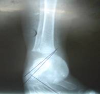 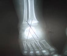
Fig. 2(a) Fig. 2(b)
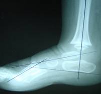 
Fig. 2(c) Fig. 2(d)
Fig. 2(a) & 4(b): Shows pre & post operative X – rays on A-P view respectively.
Fig. 4(c) & 4(d): Shows pre & post operative X - rays on Lateral view respectively.
Discussion:
The complex three dimensional deformities in a rigid, neglected or relapse clubfoot often needs to be treated by various surgical procedures. In spite of so many advances in the knowledge and techniques, the clinical outcome of neglected, resistant and relapsed or recurrent clubfoot has not changed substantially.
In our series, pre operatively, ankle motion and hind foot appearance was unsatisfactory in all cases in both the groups. Post operatively, both these parameters showed marked improvement in both the groups but still fell short of normal. Hind foot appearance was satisfactory in 93.33% feet in group II, as compared to 78.125% feet in group I. Similarly 86.67% feet had satisfactory ankle motion in group II, as compared to 68.75% feet in group I. Laaveg and Ponseti4 (1980) also observed that in majority of feet, foot and ankle motion was limited after surgery. Adelaar and Kyles5 (1981) noted that dorsiflexion is difficult to achieve, particularly if anatomical deformity is present in talus and so the result is compromised.
Patients treated by JESS distractor had better clinical results (76.67% satisfactory) than those treated by PMSTR (68.75%), even though the mean age in JESS group was much higher than PMSTR Group. Our results are comparable with the results obtained by other authors. Turco6 (1979) used clinical criteria and reported 83.8% excellent and good results using PMSTR. Mc Kay7 (1983) used clinical criteria and reported 81.8% excellent and good results. Yamamoto8 (1988) used clinical criteria and reported 70.4% excellent and good results after performing PMSTR. Simons9 (1985) after performing complete subtalar release and using same criteria found 72% cases with satisfactory clinical results. Grill and Frankie10 (1987) achieved plantigrade foot with satisfactory clinical and radiological results in all 10 feet treated using Illizarov technique. S Suresh et al11 used clinical criteria and reported 91% excellent and good results using Jess fixator.
Preoperatively radiological assessment revealed that lateral talocalcaneal angle (which is an indicator of hindfoot varus) and tibiocalcaneal angle (which is an indicator of hindfoot equinus) were unsatisfactory in all feet. However some feet which had received regular conservative treatment had normal anterior talocalcaneal angle (indicator of varus angulation of hindfoot). None of these feet had a satisfactory lateral talocalcaneal angle or tibiocalcaneal angle. Atttenborough12 (1966) had also observed that manipulation and cast treatment often failed to correct equinus and produced false corrections. Sudmann et al13 (1983) stated that equinus is the most resistant component of clubfoot deformity and that tendoachilles and tibialis posterior are the main dynamic factors responsible for resistance to correction.
The anterior talocalcaneal angle was taken as an indicator of hindfoot varus. Following surgery it was satisfactory in all feet (100%) in group I and group II. Lateral talocalcaneal angle (indicator of hindfoot varus) was more often satisfactory in group II (80%) as compared to group I (68.75%). Calcaneus – first metatarsal angle (which is an indicator of cavus deformity) was in satisfactory range in both the groups, group I (84.375%) and group II (83.33%). Calcaneus – second metatarsal angle (which is an indicator of forefoot adduction) was in satisfactory range in 22 cases of group II (73.33%) as compared to 22 cases of group I (68.75%). Similar observations were found in reference to the tibiotalar angle (indicator of equinus at hindfoot) which was satisfactory in 22 cases (73.33%) of group II and 21 cases of group I (65.625%). However it was observed that all angles were corrected to a greater extent in group II as compared to group I.
In both the groups, the deformity most commonly left unsatisfactory, both clinically and radiologically ( calcaneo second metatarsal angle) was forefoot adductions. A number of authors have also reported forefoot adduction as most common residual deformity eg. Cohen – Sobel et al14 (1993) 60%, Ponseti I V15 (1992) 48%.
In group I the results were clinically and radiologically not satisfactory in any of the case when the age for carrying out PMSTR was more than 12 months whereas it was satisfactory in 72.51% cases if the surgery was performed before 12 months of age. This suggested that ideal time for carrying out PMSTR is before12 months of age.
Various authors have reported better results if soft tissue surgery is done at a younger age. Turco6 (1979) reported best results in the age group of 1-2 years.
In group II the youngest child was 11 months old and the oldest was of 4 years of age. It was found that patients undergoing JESS surgery with age less than 2 years had a very low complication rate (13.33%) compared to patients who were more than 2 years of age. Common complications in this group were : pin tract infections 4 cases(13.33%); pressure sore( due to foot plate)causing superficial necrosis of plantar surface of toes in 7 cases(23.33%); and excessive valgus in 3 patients ( 10 % ). Thus, JESS procedure offers significantly better results if surgery is done before two years of age in such cases.
Our results are slightly lower as compared to other authors6,7,10,11, probably because all of them used mainly clinical criteria and taleocalcaneal index to assess result after surgery, while we used a much more rigid clinical and radiological criteria.
Conclusion:
The best treatment for neglected, resistant and relapsed or recurrent clubfoot remains controversial. In absence of a universally acceptable method of deformity assessment, results of various studies cannot be compared.
Based on our experience we recommend that PMSTR offers best results in rigid or relapsed cases of CTEV when the surgery is performed within 1 year of age as there are good chances of achieving the goal of obtaining a cosmetically acceptable, pliable, functional, painless, and plantigrade foot. After the age of 1 year, results are compromised, as adequate reduction is difficult to achieve due to soft tissue and bony changes.
However, JESS is ideally suited for a case of neglected, resistant and relapsed or recurrent clubfoot when such a child reports late. Better results are obtained if Jess treatment is initiated before two years of age. However, even in age group of 2 to 4 years, it offers good clinical and radiological outcome. JESS is technically a bit complicated procedure and requires motivated parents to adhere strictly to the distraction protocol and subsequent follow up for good results.
Reference :
- Hippocrates(400B.C.): The genuine work of Hippocrates translated from Greek by Francis Adams with an introduction by E.C.Kelly. Baltimore: Williams& Wilkins,1939.
- Ilizarov GA. Clinical application of the tension-stress effect for limb lengthening. Clin Orthop 1990; 250:8–26.
- Joshi, B.B.; Laud, N.S.; Warrier, S.S. Kanaji BG, Joshi AP, Dabake H: Treatment of CTEV by Joshi External Stabilization System (JESS) in : Kulkarni GS, Editor. Textbook of Orthopedics and Trauma, Ist Edition, New Delhi: Jaypee BrothersMedical Publihers Ltd; 1999.
- Laaveg, S.J Aand Ponseti,I.V.; Long term results of treatment of congenital club foot: J.Bone Jt Surgery(1980), 62A,23.
- Adelaar, R.S.and Kyles, M.K.; Surgical correction of resistant talipes equinovarous- Observations and Analysis. Apreliminary Report: Foot and Ankle(1981) 2 (3),126.
- Turco, V.J.: Resistant congenital club foot- One stage posteromedial release with internal fixation; a follow up report of a fifteen years experience. J. Bones Jt. Surg.(1979), 61A, 805-14
- McKay, D.W.:New concept and approach to club foot treatment:Section II- Correction of club foot. J.Pediat.Orthop (1983),(2),347.
- Yamamoto, H. And FURUYA,K.(1988): One Stage Posteromedial release of club foot; J. of Paediatric Orthopaedics,(1988), Vol(8), Issue 5, 590.
Simons, G.W. Complete subtalar release in clubfeet. J. Bone Jt. Surg1985. 67A:1056-1065.
- Grill F, Franke J. The Ilizarov distractor for the correction of relapsed or neglected clubfoot. J Bone Joint Surg Br 1987; 69B: 593–597.
Suresh S; Ahmed A; SharmaVK Role of Joshi’s external stabilisation system fixator in the management of idiopathic clubfoot. Journal of Orthopaedic Surgery 2003: 11(2): 194–201
- Attenborough, C. G. : Severe congenital talipes equino various ; J. BONE Jt Surg.’ (1966), 48 B, 31-39.
- Sudmann,E., Hald, J.K. and Skandfer, B.: Feature resisting primary treatment of congenital club foot; Acta Orthop. Scand., (1983),54(6),850-857.
- Cohen-Sobel, E,; Caselli, M.; Giorgini, R. Etal: Long term follow up of club foot surgery; Analysis of 44 patients. J. Foot Ankle Surg., (1993),32(4), 411-423.
11.Ponseti, I.V.: Treatment of congenital clubfoot; J. Bones Jt Surg.(1992) ,74A; 448-454.
|

