Address for Correspondence
Yulija M. Cherniakova
Oktober Av.1, ap. 55,
Gomel,
Belarus, 246056.
E-mail: ychernyakova72@mail.ru
Abstract:
The paper presents the findings of the 2-year study of 16 patients with posttraumatic osteoarthritis of the knee joint and intolerance to hyaluronates. Intra-articular injections of doxycycline-modified blood serum were used as medication. The treatment results were compared with those of 23 OA patients medicated with the hyaluronic substitute of synovial fluid Synvisc. Joint function was assessed by the scales VAS and WOMAC and for assessment of the quality of life the health survey SF-36 was used. The effect of blood autologous serum is comparable to that of hyaluronic synovia substitute. This is attributed to the similar properties and biochemical composition of serum and synovia. The curative effect was achieved owing to improvement of lubrication and nutrition of cartilages, presence of growth factors in serum, and decreasing coefficient of friction in the joint. Chondroprotective effect of doxycycline is due to inhibited lymphocyte migration, suppression of matrix metalloproteinases in the cartilage and NO synthase. Therefore, transfusion chondroprotection is an alternative to hyaluronate treatment and can be recommended to hyaluronate-intolerant patients with OA of the knee joint.
J.Orthopaedics 2011;8(2)e7
Introduction
Posttraumatic osteoarthritis (OA) of knee joint covers 15% of all OA [1]. OA results from joint injuries happened once or manifold. OA most often develops in patients having damaged menisci and tendon breakdown resulted in discongruency of joint bone ends and instability of knee joints [2]. Though lesions of knee joints are healed using surgical technologies, OA develops with time. Pathogenesis of posttraumatic changes in the joint is due to degeneration of the hyaline cartilage, inflammation of synovial tissue, and changes in the properties and functioning of synovial fluid (SF) [3]. Tribological function of SF that consists in cartilage lubrication of a healthy joint and its minimal wear is the most essential. In traumas and inflammations of the joint, proteoglycan macromolecules of hyaluronic acid (HA) are broken down, and enzymes that damage the cartilage matrix are accumulated in SF [4].
At the initial stages of OA the preparations of hyaluronic acid are injected into the cavity of degenerated joints to provide the chondroprotection and SF substitution [5]. They protect the cartilage against axial impact mechanical loads. However, not all patients are tolerable to these drugs. In some cases reactions to injections in the form of synovitis, enhanced pains in the joint, as well as skin hyperemia and hyperthermia can happen. Such patients will soon need knee arthroplasty.
The study aims at clinical evaluation of transfusion chondroprotection as an alternative procedure in treating the patients with posttraumatic OA of knee joints and intolerance to hyaluronates.
Patients and methods
Since January, 2008 till the end of December, 2010 we were observing and treating 16 patients (group 1 – 12 male, 4 female, aged 29−46 years) with posttraumatic OA of knee joints and intolerance to hyaluronates in the Orthopaedics and Traumatology clinics at Gomel State University, Belarus. In 8 patients OA developed after meniscectomy performed more than 3 years ago and in 5 patients OA developed after intra-articular fractures of hip and/or tibia. The group included 3 sportsmen having prolonged multiple joint injuries, lesions of menisci and cruciform ligaments with signs of knee instability that were cured surgically. In single intra-articular injections of hyaluronates all the patients reported more severe pains in joints and tissue edema. In the 7 patients hyperemia and itching in the place of the injections were noted.Within the same terms we observed posttraumatic OA in 23 patients medicated only with hyaluronates. They composed control group 2. Knee joint injuries in this group were similar to the trial group and are presented in Table 1. A course of treatment in the control group consisted of three injections of Synvisk with an interval of 5−7 days and repeated in 6 months.
Patients with uncontrollable somatic pathologies (diabetes, rheumatoid arthritis, immunodepression, infections, and psychic disorders) as well as those who had intra-articular injections of hyaluronates in recent 6 months or glucocorticosteroids in recent 2 months were not included in the groups.
Table 1 Patients demographics
Description |
Group 1 (n=16), AS |
Group 2 (n=23), HA |
Gender (male:female) |
12:4 |
15:8 |
Age (years ± SD) |
37.3 ± 8.7 |
39.17± 6.3 |
Side (right:left) |
9:7 |
14:9 |
Prescription of knee injury ± SD (months) |
38.3 ± 8.4 |
36.5 ± 9.2 |
Knee injuries in anamnesis:
meniscectomy
intra-articular fracture
sports trauma, knee instability |
8
5
3 |
13
7
3 |
X-ray stage OA (I:II:III) |
4:5:7 |
11:5:7 |
Intolerance to HA |
16 |
0 |
BMI ± SD (kg/m2) |
30.4 ± 3.7 |
30.5 ± 4.1 |
AS − autologous serum, HA − hyaluronates, SD − standard deviation, BMI − body mass index
We used the blood autologous serum (AS) prepared of proper patients’ blood to treat the group 1 patients. The serum choice was due to the findings of the previous studies performed by the authors. Higher lubricity of plasma and blood serum than SF taken from osteoarthritis-damaged joint was established in vitro. Coefficients of friction of serum and normal SF are equal, namely, 0.045÷0.065 [6]. Biochemical composition of serum practically corresponds to that of normal SF because of the absence of fibrinogen found in blood. Blood serum provides the basis of SF and is a protein and electrolyte substrate that ensures cartilage trophism [7].
To enhance the chondroprotective effect, patients’ blood serum was modified in vivo with doxycycline hydrochloride. The chondroprotective effect of doxycycline is due to inhibition of polymorphic-nuclear leucocytes and blocking of the synthesis of matrix metalloproteinases in the synovial medium of joints in inflammation [8]. To modify serum, a patient took 200 mg of doxycycline one time orally. In 2 hours 30 ml of blood was taken from the elbow vein with a syringe and the syringe was incubated with no preservatives at 18−20 ºС for 5 hours. After that, 5 ml of doxycycline-saturated serum was injected with a new syringe in the knee joint. The drug was injected into the fissure of the knee joint from the outer lateral parapatellar point by directing the needle in the intercondylar space and introducing the serum in the cavity. After injection the patients were recommended to restrict loads to the joint for 2 hours. The procedure was implemented twice with an interval of 5−7 days according to the adopted technique [9]. The medication course was repeated in 6 months.
The experimental and clinical trial of transfusion chondroprotection was performed with patients’ consent observing ethical principles of Helsinki Declaration adopted on the 18th World Medical Assembly (1964), adapted to the current problems of medical experiments on the 41st World Medical Assembly in Hong Kong (1989) and in line with the rules of Good Clinical Practice (1997).
The stage of OA was determined prior to treatment using the Kellgren-Lawrence scale of the X-ray images taken in standing position, and cartilage degeneration was justified by arthroscopy. The OA stages corresponded to stages 1−3 by Kellgren-Lawrence and stages 2−4 by Outerbridge (Fig. 1). The treatment results were evaluated in 2 weeks, 3 and 6 months once each treatment course was completed in both trial and control groups. Dynamics of pain syndrome, functional state of the joint and total efficacy of the treatment were assessed using the visual analogue scale (VAS) and Western Ontario and McMaster Universities Osteoarthritis Index (WOMAC). The health-related quality of life was evaluated by means of the questionnaire of 36-Item Short-Form Health Survey(SF-36) including 8 scales (Physical Functioning – PF, Role-Physical Functioning – RP, Bodily Pain – ВР, General Health – GH, Vitality – VT, Social Functioning – SF, Role-Emotional – RE, Mental Health – MH) [10]. At the end of the second year of treatment, the “solid” final evidences were assessed, that is, the patients’ need in arthroplasty of knee joints and prolongation of the terms before surgical intervention. The control X-ray examination of the width of the knee fissure loaded by the body’s weight was performed each 6 months during the 2-year treatment.
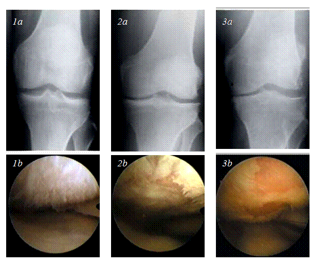
Figure X-ray and arthroscopic figures of the knee joints with different stages of OA in the patients in study: 1а, 2а, and 3а correspond to stages 1, 2, and 3 by Kellgren-Lawrence; 1b, 2b, and 3b correspond to stages 2, 3, and 4 by Outerbridge
The criteria to be analyzed were two-sided and evaluated at the 5 % level of reliability. In multiple comparisons the Bonferroni correction was applied. The groups were comparable at р>0.05. Demographic characteristics of the patients were evaluated by the Mann-Whitney’s U criterion to be used in age comparison and by the χ2 criterion to be used in patients’ distribution by gender. The OA severity was assessed during primary examination by using the Mann-Whitney’s U criterion.
The SF-36 profiles at the initial level after 2 weeks, 3, and 6 months of treatment were compared between the groups. Each index in the questionnaire SF-36 was assessed independently using the Mann-Whitney’s U criterion. In both groups the registered health state change was assessed separately using Mantel-Haenszel’s χ2 statistics. Improvement of each scale of the questionnaire SF-36 was assessed in both groups following the similar procedures. The data were calculated and processed on an IBM computer 80486 CPU 50 MHz with SAS software (version 6.08 for MS-Windows) licensed by SAS Institute Inc. (Cary, N.C.).
Results
No side effects and complications were revealed during the treatment of the patients with intra-articular injections of modified blood autologous serum. No allergic reactions to Synviskwere registered in the control group.
Dynamics of main clinical indices in the treatment of the trial group with transfusion chondroprotector and the control group treated with hyaluronate are presented in Table 2.
Table 2 Dynamics of clinical indices in the patients with OA in treatment with blood autologous serum and Synvisk
Group |
Dynamics of items (M ± SD) |
Pain by VAS, сm |
Pain by WOMAC,
scores |
Pain in palpation scores |
Pain in movements, scores |
Synovitis intensity, scores |
30-m walk, sec |
Climb 1 flight of stairs, sec |
Group 1, AS |
Before treatment |
5.9 ± 2.6 |
15 ± 4.3 |
0.9 ± 0.7 |
1.6 ± 1.3 |
1.4 ± 1.1 |
34.2 ± 10.6 |
26.3 ± 15.3 |
In 2 weeks after the first course |
3.6 ± 2.2 p<0.0001 |
13 ± 3.7
p<0.05 |
0.7 ± 0.7 p<0.05 |
1.0 ± 0.7 p<0.05 |
1.2 ± 1.1 p<0.001 |
31.4 ± 10.2 p<0.01 |
24.1 ± 12.3 p<0.05 |
In 3 months after the first course |
2.8 ± 2.5 p<0.0001 |
11 ± 2.8
p<0.01 |
0.5 ± 0.4 p<0.05 |
1.0 ± 0.9 p<0.05 |
1.0 ± 1.0 p<0.05 |
24.1 ± 9.3 p<0.01 |
18.1 ± 5.5 p<0.05 |
In 6 months after the first course |
3.6 ± 2.7 p<0.01 |
12 ± 3.1
p<0.01 |
0.6 ± 0.4 p<0.05 |
1.6 ± 0.8 p<0.05 |
1.1 ± 1.0 p<0.05 |
25.3 ± 7.6 p<0.01 |
20.5 ± 6.1 p<0.05 |
Group 2, HA |
Before treatment |
6.3 ± 2.1 |
15.8 ± 2.1 |
1.0 ± 0.9 |
1.6 ± 0.8 |
1.3 ± 1.1 |
32.8 ± 9.4 |
25.6 ± 9.6 |
In 2 weeks after the first course |
3.8 ± 1.9 p<0.01 |
14.7 ± 2.5 p<0.01 |
0.7 ± 0.9 p<0.05 |
1.3 ± 0.5 p<0,05 |
1.3 ± 0.9 p<0.05 |
31.3 ± 8,0 p<0.02 |
23.2 ± 10.8 p<0.05 |
In 3 months after the first course |
3.5 ± 1.5 p<0.01 |
13.4 ± 2.7 p<0.05 |
0.6 ± 0.5 p<0.05 |
1.1 ± 0.7 p<0.05 |
1.3 ± 1.1 p<0.05 |
24.3 ± 10 p<0.05 |
21.9± 10.1 p<0.05 |
In six months after the first course |
3.5 ± 1.9 p<0.05 |
13.7 ± 3.7 p<0.05 |
0.7 ± 0.3 p<0.05 |
1.3 ± 0.3 p<0.05 |
1.1 ± 1.0 p<0.05 |
26.1 ± 9.4 p<0.05 |
23.1 ± 8.7 p<0.05 |
p – reliability of the differences between indices before and after treatment in each group
In both groups the patients were comparable in demographic characteristics (Table 1) and severity of clinical OA manifestations (Table 2). The initial level by 8 health-related scales was also comparable (Table 3). After treatment the most significant changes were recorded in the pain index by VAS, knee joint pains in palpation and movements. A tendency to lower intensity of synovitis of knee joints is noted. The time for walking 30 meters on a smooth surface and time for climbing a flight of stairs reduced (p<0.05). However, any reliable differences between the groups under observation were not detected.
Table 3 Dynamics of the indices of quality of life of the patients with OA by the questionnaire SF-36
Scales SF-36 |
Group 1, AS (M ± SD) |
Group 2, HA (M ± SD) |
Before treatment |
In 2-year treatment |
Before treatment |
In 2-year treatment |
Physical Functioning - PF |
27.4 ± 19.7 |
47.4 ± 17.6 |
26.6 ± 15.7 |
45.8 ± 23.4 |
Role-Physical Functioning - RP |
18.7 ± 12.1 |
33.7 ± 8.1 |
15.7 ± 13.6 |
29.9 ± 10.5 |
Bodily pain - ВР |
41.8 ± 13.6 |
53.0 ± 12.9 |
39.3 ± 16.1 |
54.3 ± 18.4 |
General Health - GH |
49.3 ± 20.2 |
53.2 ± 19.5 |
46.9 ± 13.6 |
55.7 ± 21.7 |
Vitality - VT |
46.7 ± 23.3 |
51.1 ± 18.2 |
48,3 ± 24.5 |
52.5 ± 19.1 |
Social Functioning - SF |
60.6 ± 22.1 |
67.5 ± 22.8 |
58.1 ± 26.9 |
66.9 ± 25.3 |
Role – Emotional - RE |
33.3 ± 14.4 |
57.3 ± 14.9 |
33.4 ± 18.1 |
59.0 ± 19.8 |
Mental Health - MH |
56.8 ± 21.5 |
63.7 ± 23.3 |
57.3 ± 23.1 |
62.7 ± 22.6 |
Statistical differences were determined using bivariate χ2 tests (two-sided, with significance limit p<0.05)
Patients’ questioning by the SF-36 questionnaire yields the results of the assessment of quality of life within a 0 – 100 score range. The more limitations the patients experience in their everyday life, the lower scores were recorded. The lowest scores were obtained by the scales PF, RP, and RE in both groups. The highest scores before treatment were recorded in SF and MH and after treatment in ВР, GH, SF, RE and MH. Upon finishing treatment the reliable positive dynamics of the indicators of the quality of life was disclosed by all the SF-36 except the scale RP in both groups. We interpret the low scores in this scale as limitation in work or other daily activities as a result of a low emotional status. However, this scale depends to a larger degree on the environment rather than the health level. Any reliable differences between both groups in the scores of the SF-36 scales after treatment were not reported.
Assessment of the “solid” final evidences has not revealed patients’ needs in arthroplasty of knee joints. All the trial-group patients agreed continuing treatment by transfusion chondroprotection.
Discussion
Evaluation of the efficacy of OA therapy using transfusion chondroprotection by the summary estimation of the dynamics of clinical data and indicators of the quality of life evidences the positive changes in the patients’ state in the course of treatment. The results of therapy with modified autologous serum are comparable to the results of treatment with hyaluronate. Within two hours after 200 mg of doxycycline has been taken orally, 93% of the drug is bound to blood proteins and its maximal concentration 2.6 mkg/ml is set. A transport protein-doxycycline complex arises in the blood. In intra-articular injection of the modified serum, the transport complex reaches the damaged joint without being broken down and bypassing metabolically active organs and fibrously changed joint capsule. In the articular cavity doxycycline is released from the protein complex and has a therapeutic medicinal effect on the synovial medium of the joint. Instead of the pathologically changed SF containing the tissue decay products, anti-inflammatory cytokines, matrix metalloproteinases, etc., the articular cavity receives the drug saturated with proteins as carriers of biologically active substances. As a result, the trophism of the joint, as well as protein, aqueous, and saline metabolism is temporarily normalized. The lubricity of such protein-medicamental preparation is much higher than that of the pathologically modified SF. Owing to better sliding of the degenerated cartilage surfaces the pain syndrome decreases.
The medicinal effect of autologous serum is due to the following causes. First, the autoserum layer serves an additional damper between cartilages. Serum moves more easily in the anatomic channels of the joint normalizing its drainage and decelerating commissure formation owing to a large volume and lower viscosity compared with SF. Second, the low-molecular serum components diffuse easily through the ground substance layers among collagen beams and in the ground substance along the oriented proteoglycan molecules improving the trophism of metabolically active chondrocytes. The products of the broken cartilage are removed more effectively from its micropores, delivered to the synovial membrane, and eliminated by macrophages of the synovial membrane. Third, after injecting serum, concentration of inflammatory enzymes including hyaluronidases is reduced in the joint. This protects the synovial medium against enzymes and normalizes synthesis of full-grown glycosaminoglycans. Therefore, autologous serum normalizes the production of endogenous hyaluronate and restores the homeostatic state of the joint that lasts for several months.
It is believed that the stimulating effect of application of platelet-rich plasma on the reparation of cartilage in animals [11] and human joint ligaments [12] is associated with the presence of growth factors in plasma. Factors PDGF, TGF-β1, bFGF and VEGF, though in lower concentrations, are present in blood serum, which also create prerequisites for chondroprotection.
Conclusions
The study results yield the evidence of improvement in physical and psychological state of the patients with OA of knee joints treated with the transfusion chondroprotection method. Satisfactory tolerance to intra-articular injections of blood serum modified by drugs and efficacy of the treatment comparable to hyaluronic substitute injections is reported. This makes possible applying the safe and efficient therapy of OA with autologous serum for treating hyaluronate-intolerant patients.
Acknowledgments
The authors are grateful to the specialists of Orthopedics and Traumatology Clinic at Gomel State Medical University for collaboration in treating patients with OA. We also thank Professor V.A. Goldade, Head of sealing technology department, V.A. Belyi Metal-Polymer Research Institute of National Academy of Sciences of Belarus, for the opportunity to study biophysical properties of blood serum and synovial fluid.
Author contributions statement
Authors participated to an equal degree in working out of the conception of transfusion chondroprotection, assessment of treated joints functions, and the results analyzing. Dr. Yu. Cherniakova elaborated procedure of the autologous serum modification by drug and executed treatment of joints.
The authors declare that they have no conflict of interest.
References
- Sutton AJ, Muir KR, Mockett S, Fentem P. A case-control study to investigate the relation between low and moderate levels of physical activity and osteoarthritis of the knee using data collected as part of the Allied Dunbar National Fitness Survey. The Ann of the Rheum Dis. 2001; 60: 756-764.
- Thelin N, Holmberg S, Thelin A. Knee injuries account for the sports-related increased risk of knee osteoarthritis. Scand J Med Sci Sports. 2006; 16(5): 329-333.
- Monsour JM, Mow VC. On the natural lubrication of synovial joints: normal and degenerate. Trans ASME. 1977; 99: 163-173.
- Ed by Hascall VC and Kuettner KE. The many faces of osteoarthritis. Birkhauser Verlag: Basel-Boston-Berlin; 2002.
- Vad VB, Sakalkale D, Sculco TP, Wickiewicz TL. Role of the hilan G−F20 in treatment of osteoarthritis of the hip joint. Archive Phys Med Rehabil. 2003; 84: 1224-1226.
- Chernyakova YuM, Kadolich ZhV, Pinchuk LS, et al. Effect of electromagnetic field on the tribological behavior of synovial fluid. Journal of Friction and Wear. 2003; 24(6): 636-641.
- Pavlova VN. Synovial Medium of Joints. Medicine: Moscow; 1980.
- O'Dell JR, Hairy CE, Palmer W, et al. Treatment of early rheumatoid arthritis with minocycline or placebo: results of a randomized, double-blind, placebo-controlled trial. Arthritis Rheum. 1997; 40: 842-848.
- Boltrukevich SI, Chernyakova YuM, Karev DB, Pinchuk LS. Instruction N 58-0609 A method for intra-articular chondroprotection of joints with blood autoserum modified by doxycycline. Belarus, Ministry of Health; 2010; 3(11): 139-146.
- Ware JE, Kosinski M, Keller SD. SF-36 Physical and Mental Health Summary Scales: A User's Manual Boston MA: The Health Institute; 1994.
- Sun Y, Feng Y, Zhang CJ, et al. The regenerative effect of platelet-rich plasma on healing in large osteochondral defects. Int Orthop. 2010; 34(4): 589-597.
- Filardo G, Kon E, Villa SD, et al. Use of platelet-rich plasma for the treatment of refractory jumper's knee. Int Orthop. 2010; 34(6): 909-915.
|


