| ORIGINAL
ARTICLE |
|
Unstable Intertrochanteric Fracture
In Elderly Patients Bipolar Arthroplasty Or Internal Fixation?A
Matched Pair Analysis Of High Risk Cohort To Compare Mortality And
Morbidity In Two Group |
|
Patil
Suresh S*, Panghate Atul**
*Senior Registrar
**Lecturer, Dept. of Orthopaedics
Department Of Orthopaedics, 6th Floor M.S.B., K.E.M.
Hospital, Parel, Mumbai-400012
Address for Correspondence:
Patil Suresh
S.
Department Of Orthopaedics,
6th Floor M.S.B., K.E.M. Hospital,
Parel, Mumbai-400012
TelephoneNo:+91-9322952723
E-Mail :drsureshspatil@gmail.com
|
|
Abstract:
Aim: To
compare the mortality and morbidity and post op complication in
high risk Intertrochanteric fractures treated by cemented
bipolar and internal fixation.
Material
and methods:
Thirty five selected patients matched for age, sex, weight,
fracture type and preop ASA grade-III were treated by primary
bipolar arthroplasty (Group A) from January 2002 to June 2005.
All patients were operated by the same surgeon. Bipolar implants
were cemented (tapered design, 2nd generation
cemented technique, standard length) and trochanteric
comminution was circlage to restore abductor mechanism The
results of this group of patients were compared with thirty nine
patients treated with Internal Fixation (Group B) by Dynamic Hip
Screw and side plate with appropriate reduction and fixation.The
comparison was done with emphasis on perioperative mortality and
morbidity in terms of day of full weight bearing, pressure sore, pulmonary complication.
Discussion
and result:
The Bipolar Arthroplasty (Group A) was able to full weight bear
significantly earlier than the Internal Fixation (Group B)
patients. Rehabilitation was easier and faster and
post op morbidity like pressure sore pulmonary complication was
significantly low (P<0.05). The mortality in cemented bipolar
group (5/35) was significantly lower than internal fixation
group (12/39) (P<0.05).
Conclusion:
Statistically
Mortality and morbidity in bipolar Arthroplasty (Group A) was
significantly lower compared to internal fixation (Group B).
Bipolar Arthroplasty may be a better alternative treatment for
osteoporotic unstable Intertrochanteric fractures in elderly
morbid patients.
J.Orthopaedics 2008;5(3)e7
Keywords:
fracture fixation, internal; hip fractures; hip prosthesis;
Introduction:
Intertrochanteric fractures are major cause of disability
and death in elderly. The incidence of all hip fractures is
approximately 80 per 100,000 persons and is expected to double
over the next 50 years as the population ages
(1).Intertrochanteric fracture make up 45% of all hip
fractures.
Unstable intertrochanteric fractures in elderly patients are
associated with high rates of morbidity and mortality (2)
although the results have improved with the use of internal
fixation. In these patients however, comminution,
osteoporosis, and instability often preclude the early
resumption of full weight bearing (3).Treatment with primary
bipolar arthroplasty rather than internal fixation could
perhaps return these patients to their preinjury level of
activity more quickly, thus obviating the postoperative
complications caused by immobilization or failure of the
implant (4).
Recent publications indicate concern with excessive sliding of
these fixation devices when used in unstable intertrochanteric
fractures (5).The excessive sliding can result in unacceptable
shortening and external rotation deformity of the limb. Bendo
et all
reported that
most of the patients with moderate or severe collapse had poor
functional results. Elderly patients often are unable to
cooperate with partial weight bearing, or if allowed full weight
bearing, voluntarily limit loading of the injured limb (6). To
allow immediate postoperative full weight bearing and to avoid
excessive collapse at the fracture site, some surgeon (7)
recommended prosthetic replacements for unstable
intertrochanteric fracture
The
purpose of this study was to determine whether cemented
hemiarthroplasty using a standard femoral stem is a reasonable
alternative to reduction and fixation with sliding hip screws
for elderly patients in unstable intertrochanteric fracture to
reduce mortality and morbidity in term of day of full weight
bearing, pressure sore,
pulmonary complication and deep vein thrombosis associate with
long rehabilitation
Material and Methods :
Between Jan 2002 and Nov 2005 Seventy four patients who were
older than sixty five years, associated with preexisting
systemic disease, who are high risk for anaesthesia (ASA Grade
III &IV),osteoporosis as asses by Singhs index and who had been
independently mobile before sustaining an unstable
intertrochanteric fracture were treated by the same surgical
team
Patients who were unable to walk before the fracture, who were
younger than sixty five years old, not associated with any
medical disease or who had stable fracture with intact lesser
trochanter been not included in the study
Table.1 Properative Clinical Data Of Seventy Four
Patients Who Had An Unstable Intertr0chanteric
Fracture
Treated By Primary Bipolar
Arthroplasty
Or Internal Fixation With Dhs And Side Plate
|
|
Group A
|
Group B
|
|
Number of
patient |
35 |
39 |
|
Age
Sex male
female |
>65yrs
12
23 |
>65yrs
14
25 |
|
Disease
preoperatively
Cardiovascular
Chronic Lung disease
Renal
disease
Liver
dysfunction
Hypertension
Diabetes
|
14
5
2
3
4
7
|
16
5
3
4
4
7
|
Bipolar
Arthroplasty (Group-A)
This group
consist of 35 consecutive patient (23 womens) underwent
primary cemented bipolar arthroplasty. Preoperatively fourteen
patient( 40%) had cardiovascular disease; five (14% ),a
chronic lung disease; two( 6% ), a renal disease; three( 8.5%
), a liver dysfunction; four(11.5% ), a hypertension;
seven(20% ) ,a diabetic(Table-1)
A
standard length stem (small-133mm, standard-138mm,
large-143mm), tapered design femoral component with modern
technique of cementing used; greater trochanter fixed to the
proximal part of femur and to lateral part of prosthesis by
two TBW wires, lesser trochanter fixed to medial part with
circlage wire passed through hole, self centering cup is
available with 7 different sizes with external diameter ranges
from 39 to 51 millimeter.
The
appropriate templates are placed over the preoperative
roentgenograms of the fractured femur and the contralateral,
uninvolved femur in order to determine the outer diameter of the
acetabular component, and the length of the extramedullary part
of the femoral component that is needed to achieve equal limb
lengths. The final size of the prosthetic components is chosen
intraoperatively, on the basis of the desired tension in the
abductor muscles of the hip as determined during the last trial
reduction
Surgical
Technique:
The
patient is placed in lateral decubitis position on a standard
operating table, with the trunk supported by chest-rests. The
surgical approach is through a standard posterior incision, the
subcutaneous layer is dissected and the fascia lata is split
longitudinally, exposing the fracture hematoma and the greater
trochanter. superior part of greater trochanter attached to
abductors are reflected superiorly and head approached through
the fracture site without cutting external rotators, The outer
diameter of the self-centering cup that is to be used is
determined by measuring the diameter of the resected .
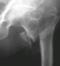
Fig.1-A
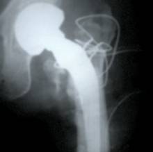
Fig.1-B
Fig. 1-A:
A seventy year-old patient who had a comminuted trochanteric
fracture.
Fig. 1-B:
Immidiate postop Radiograph, shows cemented bipolar arthroplasty
with
Wiring of the lesser and the greater trochanter
femoral head,
after exposing the proximal femoral diaphysis, canal prepared
the femoral shaft is then prepared first by a straight
intramedullary reamer is used, and next the proximal part of the
shaft is prepared further with a reamer that has a conical
enlargement, so that a correct fit is obtained between it and
the larger proximal shaft of the prosthesis. The appropriate
length of the extramedullary portion of the femoral component
can be chosen by using the adjustable trial stem. The trial stem
is assembled with a trial cup, and test reductions are performed
to determine the exact length that will provide the desired
tension of the abductor muscles. The hip is dislocated again,
and all trial components are removed. Before cementing two wires
passed in proximal diaphysis so that we can use for subsequent
reattachment of greater trochanter and a circlage wire is passed
through the lesser trochanter to permit its subsequent fixation
to the medial side of the femoral component. The femoral stem
was cemented in place using standard modern cementing techniques
that include lavage, cleaning, drying, and plugging of the
canal. After all components have been removed, a cement
restrictor is inserted and the medullary canal is rinsed with
saline solution. One or two units of polymethylmethacrylate
cement (CMW-3) are injected under pressure, and the femoral
component after the polymethylmethacrylate has set, the
self-centering cup is locked onto the prosthetic head and the
prosthesis is into the acetabulum. The two wires that we passed
previously in proximal diaphysis are passed through greater
trochanter and gradual tightening done so as to approximate
fracture site and third wire encirclaging the proximal diaphysis
that we previously positioned through lesser trochanter .The
fascia lata is closed and sutured, and the skin is closed.
Suction drainage is used in all patients for forty-eight hours.
An antibiotic is given just before operation and is continued
for five days postoperatively.
Postoperatively, the patients receive thromboembolic
prophylaxis (LMW). Unless contraindicated, anti-inflammatory
medication is administered postoperatively for one week Active
and passive mobilization of both limbs is started as soon as
possible, taking care to avoid forced adduction or rotation of
the hip that was operated on. Moderate flexion of both the hip
and knee joints, with a large pillow between the ankles during
bed rest, is recommended. In our series, walking with full
weight-bearing was allowed on 2nd day
postoperatively. The average time to walking with full
weight-bearing was fifth days.
Roentgenograms:
That
were made at three, six, and twelve months, two year
postoperatively were analyzed. The duration of follow-up
ranged from six months to three years, with an average of
eighteen months; it was determined largely by how long the
patients lived, as most of them were quite elderly.
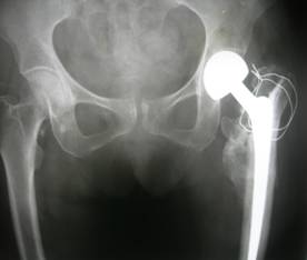
Fig.2-A
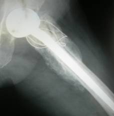
Fig.2-B
Roentgenograms mad at two years (Fig.2-A, anteroposterior &
Fig.2-B, lateral View) showing good bone formation around
lesser and greater trochanter .
Internal
Fixation
(Group B)
This group
consist of 39 consecutive patient (25 womens) underwent
Internal fixation with DHS with side plate. Preoperatively
sixteen patient(41%) had cardiovascular disease; five (13%),a
chronic lung disease; three(7.6% ), a renal disease;
four(10.2%), a liver dysfunction; four( 10.2%), a hypertension;
seven(18%) ,a diabetic (Table-1)
Surgical
Technique:
The
operation is performed on an orthopaedic fracture table, with
the patient lying supine. Fluoroscopy is routinely used. The aim
of the closed reduction is to obtain
An optimum
position, with a correct angle between the femoral neck and
shaft193839. The proximal part of the femur is exposed
through a lateral approach383948, and DHS with side plate is
inserted. Postoperatively, the patients receive thromboembolic
prophylaxis (LMW) unless contraindicated and analgesic, Sitting
up in a chair and walking without weight bearing on the
fractured limb are permitted as soon as possible.
The operating
surgeon determines when the patient should start walking, on the
basis of the stability of fixation at operation and the findings
on the postoperative roentgenograms. Non-weight-bearing is
continued until consolidation is confirmed roentgenographically.
Full weight-bearing is allowed only when complete osseous
healing has occurred. The average time from operation to walking
with full
Weight-bearing
without support was 3.5 months. Prophylactic antibiotics
were given for five days routinely. And roentgenograms of the
fractured hip were made, both at regular intervals, until the
fracture had united or technical failure had occurred. Technical
failure
Was defined as
the absence of fracture-healing, with breakage of the implant
that necessitated reoperation.
Results :
Analysis of
result by using chi-square test, unpaired t test, Fisher exact
test
There were no
significant differences between the two groups in terms of
demographic data (age, sex), fracture type, preoperative Singhs
index, ASA Grade for anaesthesia and preoperative systemic
disease (Table 1)
Also
there were no significant difference (p >0.05) between operative
time, blood loss, and hospital stay (Table2)
Table-2
|
|
Group A
|
B Group
|
p value |
|
Operative
time |
110 min
|
102 min
|
>0.05
|
|
Blood loss
|
420 ml
|
450 ml
|
>0.05
|
|
Hospital
stay
|
15 days |
18 days |
>0.05 |
The time to
full weight bearing was significantly earlier in patients who
underwent hemiarthroplasty; the mean follow-up period for the
hemiarthroplasty group was 24 months (range, 636 months). Five
(14%) of the 35 patients died in the first half year. Among
them, one developed deep infection on day15 and did not respond
to postoperative antibiotics. One had a pulmonary infection and
one sustained a cerebral hemorrhage (associated with
hypertension) both within one month. The remaining 2 patients
died from causes unrelated to the primary injury. Among the 30
patients still surviving, early complications included 2 with
bed sores,1 had pulmonary infection and 1 had intraop fracture
of proximal femoral diaphysis at time of preparation of canal
due narrow canal. Two patients were unable to walk
because of unrelated conditions. There was no dislocation,
apparent acetabular protrusion or aseptic loosening of the stem.
Require long term follow up to asses these complication
The
internal fixation group fitted with a DHS was followed up for a
mean of 23 months (range, 638 months). Twelve (31%) of the 39
patients died in the first half year; one sustained a cerebral
infarct from thromboembolism after 2 months. The remaining 11
deaths were attributed to pre-existing systemic disease. Six
months after surgery, 27 (69%) of the 39 patients in this group
were surviving Among them, 18 developed early complications ;bed
sores in 9,pulmonary infection in 5,mechanical failure in 4 who
underwent revision surgery by arthroplasty or implant removal.
Discussion :
Unsatisfactory
surgical outcome is common in elderly patients with
intertrochanteric fractures; medical illness, osteoporosis, and
fracture instability are contributing factors. Early
mobilisation may decrease the risk of mortality and morbidity,
although older patients are unable to walk well and only capable
of partial weight bearing in the postoperative period. (8)
In
patients with osteoporotic fractures, maintenance of reduction
can be a major problem during the healing period. To reduce the
healing time, dynamic devices are replaced with the static ones.
Biomechanical studies show that dynamic implants have more
weight-bearing capacity than static implants.(9)Furthermore,
partial weight bearing creates a micromovement in the dynamic
systems which increases union rate. However, cut-out is the main
complication of internal fixation. Central positioning of the
screw in the femoral neck has been recommended, (10) which
yields cut-out rate of about 13%. The strength of fixation
depends on screw positioning and bone quality. The cut-out rate
in the present study was 10% and the respective patients
underwent revision surgery (arthroplasty or implant removal).
Many
surgeons prefer arthroplasty for the treatment of unstable
trochanteric fractures in the elderly in order to decrease
complications: Rosenfeld et al.(11) used arthroplasty and
reported 86% satisfactory results in the early period. Stern and
Angerman9 reported 94% good and excellent results after a mean
follow-up period of 8 months. Haentjens et al. (12) compared the
clinical results of internal fixation and bipolar arthroplasty
for unstable trochanteric fractures and reported 75%
satisfactory results and less postoperative complications in the
latter group. They insisted that early weight bearing was the
major factor responsible for decreasing postoperative
complications.
K.casey Chan and Gurdevs.Gill (13) found that Use of
standard cemented hemiarthroplasty is a reasonable alternative
to a sliding screw device for the treatment of intertrochanteric
fractures to achieve less postoperative complication. Prof.
Chris Grimsud,Raul J. Monzon(14) treated all unstable three and
four part hip fractures with standard femoral stem and circlage
cabling of trochanters and they conclude that bipolar
arthroplasty allows safe early weight bearing on the injured
hip and had a relatively low rate of complication
P. Florian
Geiger; P.Monique Zimmermann-Stenzel found that Mortality was
significantly influenced by Age, Gender, Amount of
Co-morbidities but not by fracture classification. (15)
Mortality rate of bipolar arthroplasty and internal fixation of
different study compare with current study are shown in Table
4
TABLE-4
Journal of
arthroplasty-April2005
Chris Grimsud,
Raul J. Monzon
Bipolar
Arthroplasty
|
|
MORTALITY
AT 1 yr |
|
Stern et
al
Green et
al
Chris
Grismud
Harwin et
al
Haentgens
et al
Chan et al
Current
study |
14%
20%
10.3%
NR
35%
7.3%
14% |
Internal Fixation
|
|
MORTALITY
AT 1 yr |
|
Haentgens
et a
Kyle et al
Hardy et
al
Haidukewvch
Current
study |
24%
NR
35%
19%
31% |
Bipolar arthroplasty group had a lower postoperative
complication rate and resulted in earlier weight bearing, which
was also reported by others. There was a significant difference
in full weight bearing time between the 2 groups. Though more
costly, bipolar arthroplasty is a treatment option for patients
with unstable Intertrochanteric fractures, which can achieve
earlier mobilisation.
Reference :
-
JD Zuckerman: Hip fracture. N Engl J Med 1996; 334:1519-1523.
-
Jensen, J.
S.: Trochanteric Fractures. An Epidemiological, Clinical and
Biomechanical Study. Acta Orthop. Scandinavica, Supplementum
188, 1981.
-
Bergman, G. D.; Winquist, R. A.; Mayo, K. A.; and
Hansen, S. 1. , JR. : Subtrochanteric Fracture of the
Femur. Fixation Using the Zickel Nail. J. Bone and Joint Surg., 69-A: 1032-1040, Sept. 1987.
-
Asencio, G.: La grande prothse
epiphyso-metaphyso-diaphysaire de lextremitymit suprieure du
femur de Vidal-Goalard.
Etude
clinique a propos de
265 CAS, pp. 23-29, 77-83.
Montpellier, Dehan, 1978.
-
Bendo JA,
Weiner LS, Strauss E, and Yang E: Collapse of intertrochanteric
hip fractures fixed with sliding screws. Orthop Rev Suppl:
30-37, 1994.
-
Koval KJ,
Sala DA, Knmmer FJ, Zuckerman JD: Postoperative weight-bearing
after a fracture of the Femoral neckor an intertrochanteric
fracture. J Bone Joint Surg 80A:352-356, 1998
-
Broos PL,
Rommens PM, Deleyn PR, Geens VR, Stappaerts KH: Pertrochanteric
fractures in the elderly: Are there indications for primary
prosthetic replacement? J Orthop Trauma 5:446-451, 1991
-
Leung KS,
So WS, Shen WY,
Hui PW. Gamma nails and dynamic hip screws for peritrochanteric
fractures. A randomized prospective study in elderly patients. J
Bone Joint Surg Br 1992; 74:34551.
-
Esser MP,
Kassab JY, Jones DH. Trochanteric fractures of the femur. A
randomized prospective trial comparing the Jewett nail-plate
with the dynamic hip screw. J Bone Joint Surg Br 1986;
68:55760
-
Davis TR,
Sher JL, Horsman A, Simpson M, Porter BB, Checketts RG.
Intertrochanteric femoral fractures. Mechanical failure after
internal fixation. J Bone Joint Surg Br 1990; 72:2631.
-
Rosenfeld RT, Schwartz DR,
Alter AH. Prosthetic replacement for trochanteric fractures of
the femur. J Bone Joint Surg Am 1973; 55:420.
-
Haentjens
P, Casteleyn PP, De Boeck H, Handelberg F, Opdecam P. Treatment
of unstable intertrochanteric and subtrochanteric fractures in
elderly patients. Primary bipolar arthroplasty compared with
internal fixation. J Bone Joint Surg Am 1989; 71:121425.
-
K.Casey
Chan, Gurudev s. Gill. Cemented Hemiarthroplasty for Elderly
Patients with Intertrochanteric Fractures Clinical Orthopaedic
and Related Research Number 371, pp. 206-215
-
C. Grimsrud, R. Monzon, J. Richman,
M. Ries. Cemented Hip Arthroplasty With a Novel Circlage
Cable Technique for Unstable Intertrochanteric Hip Fractures
The Journal of Arthroplasty, Volume 20, Issue 3, and Pages 337 -
343.
-
P. Florian
Geiger; P.Monique Zimmermann-Stenzel. Mortality was
significantly influenced by Age, Gender, and Amount of
Co-morbidities but not by fracture classification. Arch Orthop
trauma Surg.2007-SEPT
|
|
This is a peer reviewed paper Please cite as
:
Patil
Suresh S:
Unstable Intertrochanteric Fracture In Elderly Patients
Bipolar Arthroplasty Or Internal Fixation?A Matched Pair
Analysis Of High Risk Cohort To Compare Mortality And Morbidity
In Two Group
J.Orthopaedics 2008;5(3)e7
URL:
http://www.jortho.org/2008/5/3/e7 |
|
|

