|
Abstract
Objectives : Quantitative ultrasound
of the calcaneus is an alternative to dual-energy X-ray
absorptiometry for measurement of bone mass. Various studies
have demonstrated correlation between broadband ultrasound
attenuation and bone mineral density in Caucasian populations.
This study compares the use of broadband ultrasound attenuation
(BUA) measurements with bone mineral density (BMD) for
osteoporosis screening in the Asian population.
Design: A comparative study comparing calcaneal broadband
ultrasound attenuation measurements and hip bone mineral
density.
Materials and Methods: 32 subjects with osteoporotic hip
and vertebral fractures underwent calcaneal broadband ultrasound
attenuation and dual-energy X-ray absorptiometry. The results of
both tests are compared.
Results: There is moderate correlation with Pearsons
correlation coefficient, r = 0.688 (p<0.0001). This is
comparable to results of published series for the Caucasian
population.
Conclusions: The CUBA Clinical System is a sensitive tool
to screen for osteoporosis in the Asian population. Selective
further testing with DEXA can be performed for individuals with
low BUA measurements. This approach to screening can give
potential cost savings.
J.Orthopaedics 2006;3(1)e16
Introduction:
Since the introduction of ultrasound by
Langton, et al in 19841 for the evaluation of bone strength and
quality, ultrasound has been used for assessment of vertebral
and hip fracture risk in osteoporotic patients.2-4
Calcaneal quantitative ultrasound (QUS) is an alternative
technique to dual energy x-ray absorptiometry (DEXA) for
assessing bone mass. The QUS sonometer measures velocity of
sound (VOS, in ms-1) and broadband ultrasound attenuation (BUA,
in dB MHz-1).1 In contrast to DEXA, which solely
measures bone mineral density, QUS gives added information
regarding the microstructural properties of cancellous bone.1,5-8
While BMD accounts for a significant proportion of bone
strength,9,10 these microarchitectural properties of trabecular
bone, such as strut number and thickness, trabeculae
connectivity, orientation and spacing, are also important for
determination of fracture risk.11,12 Various studies
have shown BUA to be moderately correlated with BMD in the
Caucasian population,13-15 and it is a more significant
predictor of fracture risk compared to VOS.2,12,16,17
Osteoporotic bone, being less dense, absorbs less sound,
resulting in a reduced attenuation. Normal bone, on the other
hand, will have higher attenuation. For every decrease in BUA
of 1 standard deviation, the risk of hip fracture doubles.12,16-18
This study examines the correlation between
BUA and BMD and the potential use of CUBA clinical system as a
screening tool for osteoporosis in an Asian population.
Material and Methods :
In a separate study by Lim et al., 602
healthy Asian subjects of Chinese, Malay, Indian and Filipino
descent had QUS performed. Left calcaneal BUA was measured with
the Contact Ultrasound Bone Analyser (CUBA) clinical system,
McCue Ultrasonics Ltd (Winchester, UK).19 Acoustic coupling was
achieved with the use of silicone pads to increase contact area,
and ultrasound gel to minimize air gaps. The study established
normative data for BUA in an Asian population.
TABLE 1
|
Case |
Age |
Gender |
Heel BUA
T-score
|
Hip BMD T-Score |
-
|
82 |
F |
-2.38 |
-0.94 |
-
|
76 |
F |
-2.80 |
-1.60 |
-
|
79 |
F |
-3.94 |
-2.75 |
-
|
89 |
M |
-2.56 |
-1.63 |
-
|
62 |
F |
-2.55 |
-1.78 |
-
|
72 |
F |
-3.58 |
-2.92 |
-
|
76 |
F |
-2.17 |
-1.58 |
-
|
85 |
F |
-3.87 |
-3.42 |
-
|
67 |
F |
-3.08 |
-2.70 |
-
|
73 |
F |
-2.25 |
-1.94 |
-
|
74 |
M |
-2.01 |
-1.71 |
-
|
74 |
F |
-2.55 |
-2.31 |
-
|
63 |
F |
-3.76 |
-3.54 |
-
|
90 |
F |
-2.14 |
-2.13 |
-
|
75 |
F |
-1.90 |
-2.00 |
-
|
74 |
F |
-2.11 |
-2.21 |
-
|
90 |
F |
-2.43 |
-2.54 |
-
|
68 |
F |
-2.15 |
-2.39 |
-
|
75 |
F |
-0.90 |
-1.28 |
-
|
91 |
F |
-2.15 |
-2.67 |
-
|
92 |
F |
-2.20 |
-2.79 |
-
|
95 |
F |
-3.99 |
-4.63 |
-
|
85 |
M |
-2.88 |
-3.62 |
-
|
88 |
F |
-2.34 |
-3.08 |
-
|
65 |
F |
-1.64 |
-2.40 |
-
|
78 |
M |
-1.90 |
-2.71 |
-
|
64 |
M |
-3.13 |
-4.09 |
-
|
70 |
M |
-2.37 |
-3.49 |
-
|
70 |
F |
-3.93 |
-5.11 |
-
|
89 |
F |
-2.86 |
-4.19 |
-
|
89 |
F |
-2.79 |
-4.33 |
-
|
94 |
F |
-3.99 |
-5.57 |
Thirty-two of these patients with
osteoporotic fractures of the hip and spine had additional BMD
assessment with DEXA scans of the hip. The mean age was 79 years
(range, 62 to 95 years)(Table 1), with 26 females and 6 male
subjects. The results of calcaneal BUA and hip BMD were then
compared and the association analyzed using Pearsons
correlation coefficient. A best-fit regression line, y=mx + c
was then plotted to describe the relationship between BMD and
BUA values.
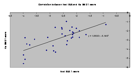
Results :
From our series, there was a moderate
correlation between left calcaneal BUA and hip BMD with
Pearsons correlation coefficient, r = 0.688 (p<0.0001). This
association, displayed graphically in Figure 1, is comparable to
results of the Caucasian population.14,15 Best fit was given by
the linear equation hip BMD = 1.0025 (heel BUA) 0.1417. In the
Caucasian population, Greenspan et al showed that BUA of the
calcaneus (CUBA) is moderately correlated with DEXA of the hip,
with Pearsons product moment correlation coefficients of 0.715.
Discussion :
WHO defined osteoporosis as a systemic
skeletal disease characterised by low bone mass and
microarchitectural deterioration of bone tissue, with a
consequent increase in bone fragility and susceptibility to
fracture.20 It is desirable to identify individuals at greatest
risk of fractures, as this risk can be halved with effective
treatment.21
In the United States, the National
Osteoporosis Foundation recommends routine BMD screening for all
women above the age of 65.22 Currently, DEXA is the gold
standard for BMD measurement. However, DEXA as a population
screening tool may not be cost-effective23 and is neither
feasible in the UK,24 nor in the Asian population owing to
limited resources and cost. In addition, bone density, being a
continuous variable, is an imperfect population screening tool
as it shows a large overlap in BMD of patients with fracture and
those without.25 Clinical risk factors are another means of
screening patients, but this has shown to be a poor
discriminator of BMD of the hip and spine.26 Other means, such
as QUS, should be evaluated for a more cost-friendly
alternative.
Besides cost, other advantages of QUS over
DEXA are that these instruments are radiation-free, inexpensive,
easy to apply, and do not require dedicated office space.15,27
BMD measurements are predictive of risk of
later fractures. A large meta-analysis involving 11 prospective
cohort studies showed that measurement of BMD at any site
(including proximal radius, distal radius, hip, lumbar spine and
calcaneus) had similar predictive ability for fractures
(relative risk 1.5, 95% CI 1.4 to 1.6) for a decrease in 1 SD in
bone density, except for measurements at hip and spine, which
have better predictive ability for fractures at hip and spine
respectively.28
For the Caucasian population, BUA has a
greater population standard deviation compared with hip BMD.29
The diagnosis of osteoporosis is currently based on the World
Health Organization (WHO) definition of BMD being more than 2.5
standard deviations below the mean for a young healthy adult
woman at spine, hip or forearm.30 However, different parts of
the skeleton behave differently within a particular subject,
with different rates of bone accretion and loss at spine, hip
and heel, and may have different BMD T-score values at these
measurement sites.31 Thus the WHO criteria for BMD T-score
values determined by DEXA may not be appropriate for
interpretation of BUA because of the different anatomic site
involved, and different measurement technology.32 As a result of
this, some studies recommend the use of an adjusted BUA T-score
of 2.0 SD instead.29,33
Based on this adjusted BUA T-score reference
range, only 1 case (Subject 26) would be missed in our series,
giving a sensitivity of 96.9%.
For cases with lower BUA than BMD T-scores,
this may be attributed to differences in the mechanical
properties of calcaneal trabecular bone, which are detected by
BUA, but not by DEXA.
The limitations of this study include the
small sample size, and the inclusion of subjects with fractures
only.
Further follow-up studies are necessary to :
(1) Establish an appropriate reference
range of BUA T-scores for the Asian population, thus allowing
QUS to be used as a diagnostic tool, minimizing the need for
further testing with DEXA, and
(2) Determine if such correlation between
BUA and DEXA exists in Asian subjects without fractures.
Determine if QUS can be used to monitor
disease progression or response to therapeutic intervention.15
This will further enhance the role of QUS in the surveillance of
patients with osteoporosis.
Conclusion:
There is moderate correlation between BUA and
BMD, with a Pearsons correlation coefficient r of 0.688
(p<0.0001) in the Asian population, comparable to studies on
Caucasian subjects. Our series demonstrates that the CUBA
Clinical System is a sensitive tool for pre-screening for
osteoporosis in an Asian population. As these instruments
provide rapid measurements, do not take up dedicated office
space, and are more affordable than DEXA of the hip and spine,
they are possible options for initial pre-screening of patients
with osteoporosis.
Further assessment of bone density and
fracture risk in these individuals can then be performed with
follow-up DEXA. This approach to population screening will
provide potential cost savings.
Reference :
-
Langton CM, Palmer SB, Porter RW. The measurement of broadband
ultrasonic attenuation in cancellous bone. Eng Med 1984;13:89-91
-
Hans D, Dargent-Molina P, Schott AM, Sebert JL, Cormier C,
Kotzki PO, Delmas PD, Pouilles JM, Breart G, Meunier PJ.
Ultrasonographic heel measurements to predict hip fracture in
elderly women:the EPIDOS prospective study. Lancet
1996;348:511-14.
-
Cepollaro C, Gonnelli S, Pondrelli C, Martini S, Montagnani A,
Rossi S, Gennari L, Gennari C. The combined use of ultrasound
and densitometry in the prediction of vertbral fracture. Br J
Radiol 1997;70:691-6.
-
Scott AM, Weill Engerer S, Hands D, Duboeuf F, Delmas PD,
Meunier PJ. Ultrasound discriminates patients with hip fracture
equally well as dual energy X-ray absorptiometry and
independently of bone mineral density. J Bone Miner Res
1995;10:243-249.
-
Gluer CC, Vahlensieck M, Faulkner KG, Engelke K,
Black D, Genant HK. Site-matched calcaneal measurementsof
broad-band ultrasound attenuation and single x-ray
absorptiometry: do they measure different skeletal properties? J
Bone Miner Res. 1992;7:1071-1079.
-
Kaufman JJ, Einhorn TA. Ultrasound assessment of bone. J Bone
Miner Res 1993;8:517-25.
-
Gluer CC, Wu CY, Genant HK. Broadband ultrasound attenuation
signals depend on trabecular orientation: an in vitro study.
Osteoporos Int 1993;3:185-191.
-
Bouxsein ML, Radloff SE. Quantitative ultrasound of the
calcaneus reflects the mechanical properties of calcaneal
travecular bone. J Bone Miner Res 1997;12:839-846.
-
Beck TJ, Ruff CB, Warden KE, Scott WW, Rao GU. Predicting
femoral strength from bone mineral data: a structural approach.
Invest Radiol. 1990;25:6-18.
-
Beck TJ, Ruff CB, Warden KE, Scott WW, Rao GU. Predicting
femoral strength from bone mineral data: a structural approach.
Invest Radiol. 1990;25:6-18.
-
Hayes WC, Piazza SJ, Zysset PK. Biomechanics of fracture risk
prediction of the hip and spine by quantitative computed
tomography. Radiol Clin North Am. 1991;29:1-18.
-
Bauer DC, Gluer CC, Cauley JA, Vogt TM, Ensrud
KE, Genant HK, Black DM. Boardband ultrasound attenuation
predicts fractures strongly and independently of densitometry in
older women: A prospective study. Arch Int Med 1997;157:629-634.
-
Young H, Howey S, Purdie DW. Broadband ultrasound attenuation
compared with dual-energy X-ray absorptiometry in screening for
postmenopausal low bone density. Osteoporos Int. 1993;3:160-164.
-
Faulkner KG, McClung MR, Coleman LJ, Kingston-Sandahl E.
Quantitative ultrasound of the heel: correlation with
densitometric measurement at different skeletal sites.
Osteoporos Int. 1994;4:42-47.
-
Greenspan SL, Bouxsein ML, Melton ME, Kolodny AH, Clair JH,
Delucca PT, Stek, M Jr, Faulkner KG, Orwoll ES. Precision and
discriminatory ability of calcaneal bone assessment
technologies. J Bone and Mineral Research 1997;12:1303-1313.
-
Pluijm SMF, Graafmans WC, Bouter LM, Lips P. Ultrasound
measurements for the prediction of osteoporotic fractures in
elderly people. Osteoporosis Int 1999;9:550-6.
-
Porter RW, Miller CG, Grainger D, Palmer SB.
Prediction of hip fracture in elderly women:a prospective
study. BMJ 1990;301:638-641.
-
Marshall D, Johnell O, Wedel H. Meta-analysis of how well
measures of bone mineral density predict occurrence of
osteoporotic fractures. BMJ 1996;312:1254-9.
-
Lim YW, Chan L, Lam KS. Broadband ultrasound attenuation
reference database for Southeast Asian males and females. Ann
Acad Med Singapore 2005;34:545-3.
-
NIH Consensus Development Conference. Diagnosis, prophylaxis and
treatment of osteoporosis. Am J Med 1993;94:646-650.
-
Black DM, Cummings SR, Karpf DB, Cauley JA,
Thompson DE, Nevitt MC, et al. Randomised trial of effect
of alendronate on risk of fracture in women with existing
vertebral fractures. Fracture Intervention Trial Research Group.
Lancet 1996;348: 1535-1541.
-
Physician's guide to prevention and treatment of osteoporosis.
Washington, DC: National Osteoporosis Foundation, 1998.
-
Advisory Group on Osteoporosis,Departmentof Health, 1994. Cited
in Langton CM, Ballard PA, Bennett DK, Purdie DW. A comparison
of the sensitivity and specificity of calcaneal ultrasound
measurements with clinical criteria for bone densitometry (DEXA)
referral. Clinical Rheumatology 1997;16:117-8.
-
Fogelman I. Screening for osteoporosis. BMJ 1999;319:1148-9.
-
Ross PD, Davis JW, Vogel JM, Wasnich RD. A critical review of
bone mass and the risk of fractures in osteoporosis. Calcif
Tissue Int 1990;46:149-61.
-
Ribot C, Tremolieres F, Pouilles JM. Can we detect women with
low bone mass using clinical risk factors? Am J Med
1995;98:252-5.
-
Baran DT. Quantitative ultrasound: a technique to target women
with low bone mass for preventive therapy. Am J Med.
1995;98:48S-51S.
-
Marshall D, Johnell O, Wedel H. Meta-analysis of how well
measures of bone mineral density predict occurrence of
osteoporotic fractures. BMJ 1996;312:1254-9.
-
Frost ML, Blake GM, Fogelman I. Contact quantitative ultrasound:
and evaluation of precision, fracture discrimination,
age-related bone loss and applicability of the WHO criteria.
Osteoporos Int 1999;10:441-449.
-
World Health Organisation. Assessment of fracture risk and its
application to screening for postmenopausal osteoporosis. In:
Geneva: WHO, 1994.
-
Sahota O, Pearson D, Cawte SW, San P, Hosking DJ. Site Specific
variation in the classification of osteoporosis, and the
diagnose reclassification using the lowest individual lumbar
vertebra T-score compared with the L1-L4 Mean, in early
postmenopausal Women. Osteoporosis Int 2000;11:852-7.
-
Greenspan SL, Maitland-Ramsey I, Myers E. Classification of
osteoporosis in the elderly is dependent on site-specific
analysis. Calcif Tissue Int 1996; 58: 409-414.
-
Stewart A, Reid DM. Quantitative ultrasound or clinical risk
factors which best identifies women at risk of osteoporosis?
Br J Radiol 2000;73:165-71.
|

